Fraud Blocker Skip to main content

Microbiology Collection (#9)

Microbiology, the fascinating world of tiny organisms and their impact on our lives

Background imageMicrobiology Collection: Scanning electron micrograph of just-divided HeLa cells

Scanning electron micrograph of just-divided HeLa cells. Zeiss Merlin HR-SEM

Background imageMicrobiology Collection: Conceptual image of sperm anatomy

Conceptual image of sperm anatomy

Background imageMicrobiology Collection: Microscopic view of Bacilli bacteria

Microscopic view of Bacilli bacteria

Background imageMicrobiology Collection: Anatomy structure of neurons

Anatomy structure of neurons

Background imageMicrobiology Collection: Conceptual image of plasmodium causing malaria

Conceptual image of plasmodium causing malaria

Background imageMicrobiology Collection: Microscopic view of a stem cell

Microscopic view of a stem cell. Stem cells are biological cells found in all multicellular organisms that can divide through mitosis and differentiate into diverse specialized cell types

Background imageMicrobiology Collection: Conceptual image of centrioles

Conceptual image of centrioles

Background imageMicrobiology Collection: A blastocyst begins implanting in the wall of the uterus

A blastocyst begins implanting in the wall of the uterus

Background imageMicrobiology Collection: Microscopic view of telomeres highlighted at the tips of chromosome

Microscopic view of telomeres highlighted at the tips of chromosome. A telomere is a region of the DNA sequence at the end of a chromosome

Background imageMicrobiology Collection: Microscopic view of Anthrax

Microscopic view of Anthrax. Anthrax is an acute disease caused by the bacterium Bacillus anthracis. Most forms of the disease are lethal, and it affects both humans and animals

Background imageMicrobiology Collection: Multiphoton fluorescence image of HeLa cells

Multiphoton fluorescence image of HeLa cells with cytoskeletal microtubules (magenta) and DNA (cyan)

Background imageMicrobiology Collection: Microscopic view of streptococcus

Microscopic view of streptococcus

Background imageMicrobiology Collection: Red blood cell flow inside the artery

Red blood cell flow inside the artery

Background imageMicrobiology Collection: Conceptual image of cellulose nanorods

Conceptual image of cellulose nanorods. Cellulose consists of both crystalline and amorphous regions

Background imageMicrobiology Collection: Microscopic view of ligand binding to a receptor protein

Microscopic view of ligand binding to a receptor protein
Microscopic view of ligand binding to a receptor. The receptor protein alters its chemical conformation (three dimensional shape)

Background imageMicrobiology Collection: A colorized scanning electron micrograph of a white blood cell eating MRSA

A colorized scanning electron micrograph of a white blood cell eating MRSA
A colorized scanning electron micrograph of a white blood cell eating an antibiotic resistant strain of Staphylococcus aureus bacteria, commonly known as MRSA

Background imageMicrobiology Collection: Microscopic view of bacteriophage attacking bacteria

Microscopic view of bacteriophage attacking bacteria

Background imageMicrobiology Collection: Conceptual image of stratified squamous epithelium

Conceptual image of stratified squamous epithelium

Background imageMicrobiology Collection: Conceptual image of synapse receptors

Conceptual image of synapse receptors

Background imageMicrobiology Collection: Conceptual image of Lactobacillus acidophilus

Conceptual image of Lactobacillus acidophilus. Lactobacillus is a gram-positive bacteria that occurs naturally in the human and animal gastrointestinal tract and mouth

Background imageMicrobiology Collection: Conceptual image of human cytomegalovirus

Conceptual image of human cytomegalovirus. Cytomegalovirus is a genus of the viral family Herpesviridae

Background imageMicrobiology Collection: Microscopic view of Sindbis virus

Microscopic view of Sindbis virus (SINV). SINV is a mosquito-borne virus that causes rash and arthritis, has been causing outbreaks in humans

Background imageMicrobiology Collection: Microscopic view of cocci bacterium

Microscopic view of cocci bacterium

Background imageMicrobiology Collection: Microscopic view of H5N1 virus with red blood cells and white blood cells

Microscopic view of H5N1 virus with red blood cells and white blood cells

Background imageMicrobiology Collection: Conceptual image of male sperm

Conceptual image of male sperm

Background imageMicrobiology Collection: Conceptual image of fertilization

Conceptual image of fertilization

Background imageMicrobiology Collection: Conceptual image of salmonella typhi causing typhoid

Conceptual image of salmonella typhi causing typhoid

Background imageMicrobiology Collection: Microscopic view of sperm traveling

Microscopic view of sperm traveling

Background imageMicrobiology Collection: Conceptual image of platelets with red blood cells

Conceptual image of platelets with red blood cells
Conceptual image of platelets with white blood cells and red blood cells

Background imageMicrobiology Collection: Conceptual image of chromosomes inside the blood stream

Conceptual image of chromosomes inside the blood stream

Background imageMicrobiology Collection: Conceptual image of simple columnar epithelium

Conceptual image of simple columnar epithelium

Background imageMicrobiology Collection: Microscopic view of Mitochondria

Microscopic view of Mitochondria. Mitochondria are the cells power producers. They convert energy into forms that are usable by the cell. Located in the cytoplasmcells activities

Background imageMicrobiology Collection: Group of vibrio cholerae bacteria which causes cholera

Group of vibrio cholerae bacteria which causes cholera. Cholera is an infection in the small intestine caused by the bacterium Vibrio cholerae. The main symptoms are diarrhea and vomiting

Background imageMicrobiology Collection: Scanning electron micrograph of a human T cell

Scanning electron micrograph of a human T cell
Scanning electron micrograph of a human T lymphocyte (also called a T cell) from the immune system of a healthy donor

Background imageMicrobiology Collection: Conceptual image of sickle cell anemia with red blood cells

Conceptual image of sickle cell anemia with red blood cells

Background imageMicrobiology Collection: Conceptual image of malaria parasites within red blood cells

Conceptual image of malaria parasites within red blood cells

Background imageMicrobiology Collection: Conceptual image of a platelet

Conceptual image of a platelet

Background imageMicrobiology Collection: Microscopic view of Dentine

Microscopic view of Dentine. Dentine is the main supporting structure of the tooth and is the second hardest tissue in the body after enamel. It is 70% mineral and acellular

Background imageMicrobiology Collection: Conceptual image of a telomere showing DNA structure

Conceptual image of a telomere showing DNA structure. A telomere is a region of the DNA sequence at the end of a chromosome. Their function is to protect the ends of the chromosome from degradation

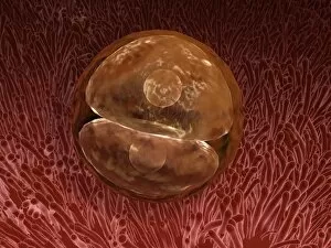
Background imageMicrobiology Collection: Zygote development 24-36 hours after fertilization

Zygote development 24-36 hours after fertilization

Background imageMicrobiology Collection: Normal artery versus artery in spasm

Normal artery versus artery in spasm

Background imageMicrobiology Collection: Conceptual image of hepatitis virus

Conceptual image of hepatitis virus. Viral hepatitis is liver inflammation due to a viral infection. The most common causes of viral hepatitis are the five unrelated hepatotropic viruses

Background imageMicrobiology Collection: Conceptual image of plasmodium

Conceptual image of plasmodium

Background imageMicrobiology Collection: Medical illustration of sperm and ovum

Medical illustration of sperm and ovum

Background imageMicrobiology Collection: Sickle cell anemia with red blood cells and white bood cells

Sickle cell anemia with red blood cells and white bood cells
Conceptual image of sickle cell anemia with red blood cells and white bood cells

Background imageMicrobiology Collection: Vials containing chemicals in research lab

Vials containing chemicals in research lab

Background imageMicrobiology Collection: Conceptual image of HIV virus

Conceptual image of HIV virus. HIV is the human immunodeficiency virus that can lead to acquired immune deficiency syndrome, or AIDS

Background imageMicrobiology Collection: Conceptual image of cytoskeleton

Conceptual image of cytoskeleton. The cytoskeleton is a cellular scaffolding or skeleton contained within a cells cytoplasm, and is present in all cells


All Professionally Made to Order for Quick Shipping

Microbiology, the fascinating world of tiny organisms and their impact on our lives. From the resilient Tardigrade or Water Bear that can survive extreme conditions to HeLa cells, a remarkable line of immortal human cells used in countless scientific studies. In Picture No. 11675590, we witness a mesmerizing light micrograph C017 / 8299 showcasing the intricate details of these microscopic wonders. The Neutrophil engulfing MRSA in SEM C018 / 8596 reminds us of the constant battle between our immune system and harmful bacteria. The beauty continues with another light micrograph C017 / 8298 capturing HeLa cells in all their glory. These cells have revolutionized medical research and paved the way for numerous breakthroughs. Yoghurt bacteria play a crucial role in transforming milk into this beloved creamy treat, highlighting how microbiology impacts even our daily diet. Stem cell research offers hope for regenerative medicine as we explore their incredible potential to repair and replace damaged tissues. Water bear once again graces us with its presence in light micrograph C016 / 8581, showcasing its unique anatomy under the microscope lens. We pay homage to Louis Pasteur, an influential figure who laid the foundation for modern microbiology through his groundbreaking discoveries and advancements in vaccination techniques. A glimpse into the invisible world reveals a microscopic view of human respiratory syncytial virus - a reminder that not all microbes are friendly companions. Embryo development captured just 24-36 hours after fertilization showcases nature's miracle unfolding before our eyes - each step meticulously orchestrated by molecular processes at work. And finally, artwork F008 /3245 portrays flu virus particles with artistic flair while reminding us of their ability to cause widespread illness if left unchecked. Microbiology unravels mysteries hidden from plain sight; it is both awe-inspiring and humbling as we delve deeper into this intricate realm where life's tiniest inhabitants shape our world.