Fraud Blocker Skip to main content

Microbiology Collection (#5)

Microbiology, the fascinating world of tiny organisms and their impact on our lives

Background imageMicrobiology Collection: PASTEUR, Louis (1822-1895) French chemist and bacteriologist. Vaccination of sheep against anthrax

PASTEUR, Louis (1822-1895) French chemist and bacteriologist. Vaccination of sheep against anthrax
Louis Pasteur (1822-1895). French chemist and bacteriologist. Vaccination of sheep against anthrax. Agerville, France, 1884. Engraving

Background imageMicrobiology Collection: Coffee under microscope

Coffee under microscope
Illustration of a coffee under microscope

Background imageMicrobiology Collection: Unicellular eukaryotic organisms

Unicellular eukaryotic organisms
Microscopic Organisms Antique Print, Vintage Lithograph, Urtiere Protozoen, Protozoa Old Zoology Illustration Unicellular Microbiology

Background imageMicrobiology Collection: Cross-section diagram of a non-cancerous tumour including a fibrous capsule

Cross-section diagram of a non-cancerous tumour including a fibrous capsule, tissue layer and blood vessel

Background imageMicrobiology Collection: Illustration of human nerve cell cross section showing dendrite, soma, axon, nucleus

Illustration of human nerve cell cross section showing dendrite, soma, axon, nucleus, nodes and myelin sheath

Background imageMicrobiology Collection: Illustration of the structure of a virus

Illustration of the structure of a virus

Background imageMicrobiology Collection: Illustration of structure of a bacterium

Illustration of structure of a bacterium

Background imageMicrobiology Collection: Illustration of immune response, involving chain of defensive white blood cells, triggered by microb

Illustration of immune response, involving chain of defensive white blood cells, triggered by microb

Background imageMicrobiology Collection: Illustration of the structure of a plant cell, including nucleus, nucleolus, ribosome, endoplasmatic

Illustration of the structure of a plant cell, including nucleus, nucleolus, ribosome, endoplasmatic

Background imageMicrobiology Collection: Cross section illustration of Synapse

Cross section illustration of Synapse

Background imageMicrobiology Collection: Cross section illustration of generalised human cell with major organelles

Cross section illustration of generalised human cell with major organelles

Background imageMicrobiology Collection: Illustration of an Eukaryote cell

Illustration of an Eukaryote cell

Background imageMicrobiology Collection: Illustration of a Prokaryote cell

Illustration of a Prokaryote cell

Background imageMicrobiology Collection: Illustration of bacterium cell reproducing

Illustration of bacterium cell reproducing

Background imageMicrobiology Collection: Illustration of different types of cells, nerve cells, red blood cell, muscle cell

Illustration of different types of cells, nerve cells, red blood cell, muscle cell, and guard cells of a leaf

Background imageMicrobiology Collection: Illustration of foraminiferan shell

Illustration of foraminiferan shell

Background imageMicrobiology Collection: Illustration of Didinium on its own and swallowing a Paramecium

Illustration of Didinium on its own and swallowing a Paramecium

Background imageMicrobiology Collection: Illustration of how an amoeba moves, liquid cytoplasm flowing through pseudopods carrying

Illustration of how an amoeba moves, liquid cytoplasm flowing through pseudopods carrying organelles with it, amoeba sending out pseudopods in direction of motion

Background imageMicrobiology Collection: Microscopic view inside of the artery with intestinal villi

Microscopic view inside of the artery with intestinal villi

Background imageMicrobiology Collection: Conceptual image of synapse of neuron inside brain

Conceptual image of synapse of neuron inside brain

Background imageMicrobiology Collection: Heart with arteries showing cholesterol in one and plaque in the other

Heart with arteries showing cholesterol in one and plaque in the other
Heart with arteries showing cholesterol in one artery and atherosclerotic plaque in the other

Background imageMicrobiology Collection: Microscopic visualization of grass pollen grains

Microscopic visualization of grass pollen grains

Background imageMicrobiology Collection: Illustration of an atypical growth on the skin that could be a sign of skin cancer

Illustration of an atypical growth on the skin that could be a sign of skin cancer
Illustration of an atypical growth on the skin that could be a warning sign of skin cancer. Sudden change in the appearance, shape, color, size

Background imageMicrobiology Collection: Researcher uses pipette in lab

Researcher uses pipette in lab

Background imageMicrobiology Collection: Single virus particle

Single virus particle

Background imageMicrobiology Collection: Conceptual image of sperm inside fallopian tube

Conceptual image of sperm inside fallopian tube

Background imageMicrobiology Collection: Red blood cells on an agar plate are used to diagnose infection

Red blood cells on an agar plate are used to diagnose infection. The plate on the left shows a positive staphyloccus infection

Background imageMicrobiology Collection: Conceptual image of antibody attaching and killing bacteria

Conceptual image of antibody attaching and killing bacteria

Background imageMicrobiology Collection: Conceptual image of a telomere

Conceptual image of a telomere. A telomere is a region of the DNA sequence at the end of a chromosome. Their function is to protect the ends of the chromosome from degradating

Background imageMicrobiology Collection: Microscopic view of histoplasmosis

Microscopic view of histoplasmosis. Histoplasmosis is an infection in the lungs caused by inhaling the spores of a fungus. This fungus, called Histoplasma capsulatum

Background imageMicrobiology Collection: Conceptual image of the Human Papilloma Virus

Conceptual image of the Human Papilloma Virus (HPV). HPV is a virus from the papillomavirus family that affects human skin and the moist membranes that line the body

Background imageMicrobiology Collection: Microscopic view of multiple nerve cells, known as neurons

Microscopic view of multiple nerve cells, known as neurons
Microscopic view of multiple nerve cells, which are also called neurons. These are responsible for passing information around the central nervous system within the human body

Background imageMicrobiology Collection: Microscopic view of diplococcus bacterium

Microscopic view of diplococcus bacterium

Background imageMicrobiology Collection: Conceptual image of cell surface receptors

Conceptual image of cell surface receptors. Cell surface receptors are specialized integral membrane proteins that take part in communication between the cell and the outside world

Background imageMicrobiology Collection: Blood flow through a relaxed artery verus an artery in spasm

Blood flow through a relaxed artery verus an artery in spasm

Background imageMicrobiology Collection: Trachea cross-section showing normal and asthmatic bronchiole

Trachea cross-section showing normal and asthmatic bronchiole
Trachea cross-section showing comparison of normal and asthmatic bronchiole

Background imageMicrobiology Collection: Rack of test tubes

Rack of test tubes

Background imageMicrobiology Collection: Illustration of muscle contraction

Illustration of muscle contraction

Background imageMicrobiology Collection: Conceptual image of cholerae bacteria

Conceptual image of cholerae bacteria

Background imageMicrobiology Collection: Microscopic view of white blood cells inside blood vessel

Microscopic view of white blood cells inside blood vessel

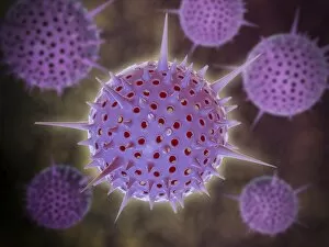
Background imageMicrobiology Collection: Conceptual image of Radiolarians with a skeletal frame

Conceptual image of Radiolarians with a skeletal frame. Radiolarians are tiny protozoans that live in the ocean

Background imageMicrobiology Collection: Conceptual image of centriole

Conceptual image of centriole

Background imageMicrobiology Collection: Microscopic view of bacteriophages on the surface of a bacteria

Microscopic view of bacteriophages on the surface of a bacteria

Background imageMicrobiology Collection: Microscopic view of a unipolar neuron

Microscopic view of a unipolar neuron. A unipolar neuron is a type of neuron in which only one protoplasmic process (neurite) extends from the cell body

Background imageMicrobiology Collection: Common cold influenza virus with microscopic close-up

Common cold influenza virus with microscopic close-up

Background imageMicrobiology Collection: Sperm traveling towards egg with cellia

Sperm traveling towards egg with cellia

Background imageMicrobiology Collection: Cancer cell with red blood cell flow

Cancer cell with red blood cell flow


All Professionally Made to Order for Quick Shipping

Microbiology, the fascinating world of tiny organisms and their impact on our lives. From the resilient Tardigrade or Water Bear that can survive extreme conditions to HeLa cells, a remarkable line of immortal human cells used in countless scientific studies. In Picture No. 11675590, we witness a mesmerizing light micrograph C017 / 8299 showcasing the intricate details of these microscopic wonders. The Neutrophil engulfing MRSA in SEM C018 / 8596 reminds us of the constant battle between our immune system and harmful bacteria. The beauty continues with another light micrograph C017 / 8298 capturing HeLa cells in all their glory. These cells have revolutionized medical research and paved the way for numerous breakthroughs. Yoghurt bacteria play a crucial role in transforming milk into this beloved creamy treat, highlighting how microbiology impacts even our daily diet. Stem cell research offers hope for regenerative medicine as we explore their incredible potential to repair and replace damaged tissues. Water bear once again graces us with its presence in light micrograph C016 / 8581, showcasing its unique anatomy under the microscope lens. We pay homage to Louis Pasteur, an influential figure who laid the foundation for modern microbiology through his groundbreaking discoveries and advancements in vaccination techniques. A glimpse into the invisible world reveals a microscopic view of human respiratory syncytial virus - a reminder that not all microbes are friendly companions. Embryo development captured just 24-36 hours after fertilization showcases nature's miracle unfolding before our eyes - each step meticulously orchestrated by molecular processes at work. And finally, artwork F008 /3245 portrays flu virus particles with artistic flair while reminding us of their ability to cause widespread illness if left unchecked. Microbiology unravels mysteries hidden from plain sight; it is both awe-inspiring and humbling as we delve deeper into this intricate realm where life's tiniest inhabitants shape our world.