Fraud Blocker Skip to main content

Microbiology Collection (#3)

Microbiology, the fascinating world of tiny organisms and their impact on our lives

Background imageMicrobiology Collection: Louis Pasteur in his laboratory

Louis Pasteur in his laboratory
Louis Pasteur (1822-1895), French chemist and microbiologist, in his laboratory. Date: 1885

Background imageMicrobiology Collection: cell division

cell division
microscopic image

Background imageMicrobiology Collection: Microscopic view of animal cell

Microscopic view of animal cell

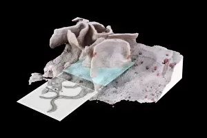
Background imageMicrobiology Collection: Histopathology and pathophysiology of diabetic food ulcers

Histopathology and pathophysiology of diabetic food ulcers

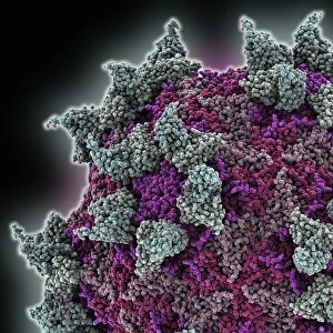
Background imageMicrobiology Collection: Rhinovirus and antibody, molecular model C015 / 7139

Rhinovirus and antibody, molecular model C015 / 7139
Rhinovirus. Molecular model of the antigen-binding fragment (Fab) from a strongly neutralising antibody bound to a human rhinovirus 14 (HRV-14) particle

Background imageMicrobiology Collection: Adenovirus hexon protein

Adenovirus hexon protein, molecular model. Hexon proteins are part of the protein coat or shell (capsid) of adenoviruses. In viruses

Background imageMicrobiology Collection: Yeast fungus skin infection, SEM

Yeast fungus skin infection, SEM
Yeast fungus skin infection. Coloured scanning electron micrograph (SEM) of human skin infected with a yeast fungus. Magnification: x3130 when printed at 10 centimetres wide

Background imageMicrobiology Collection: Louis Pasteur Experiment Rabbit Chloroformed

Louis Pasteur Experiment Rabbit Chloroformed
Associates of Louis Pasteur (left) conduct an experiment under his instructions on a chloroformed rabbit. Louis Pasteur, 1822 - 1895

Background imageMicrobiology Collection: Louis Pasteur Portrait Office Pasteur Institute

Louis Pasteur Portrait Office Pasteur Institute
Louis Pasteur in his office at the Pasteur Institute, Paris. Louis Pasteur, 1822 - 1895. French chemist and microbioligist who discovered, amongst other things

Background imageMicrobiology Collection: Louis Pasteur Portrait Laboratory French France

Louis Pasteur Portrait Laboratory French France
Louis Pasteur in his laboratory. Louis Pasteur, 1822 - 1895. French chemist and microbioligist who discovered, amongst other things, pasteurization and the principlies of vaccination

Background imageMicrobiology Collection: Louis Pasteur, 1822 - 1895. French chemist and microbiologist renowned for his discoveries of

Louis Pasteur, 1822 - 1895. French chemist and microbiologist renowned for his discoveries of the principles of vaccination, microbial fermentation and pasteurization

Background imageMicrobiology Collection: Antonie van Leeuwenhoek, 1632-1723. Dutch scientist

Antonie van Leeuwenhoek, 1632-1723. Dutch scientist

Background imageMicrobiology Collection: Clostridium tetani, causative agent of tetanus

Clostridium tetani, causative agent of tetanus
Biology - Medicine - Microbiology - Clostridium tetani, causative agent of tetanus under the microscope with contrast color

Background imageMicrobiology Collection: Visual system, Eye, Retina under microscope

Visual system, Eye, Retina under microscope
Medicine - Anatomy - Visual system - Eye - Retina under microscope

Background imageMicrobiology Collection: brain cells

brain cells
microscopic image

Background imageMicrobiology Collection: Robert Koch (1843-1910). Engraving. Colored

Robert Koch (1843-1910). Engraving. Colored
Robert Koch (1843-1910). German physician. Nobel Prize, 1905. Engraving in The Spanish and American Illustration, 1890. Colored

Background imageMicrobiology Collection: PASTEUR, Louis (1822-1895). Vaccination of sheep against ant

PASTEUR, Louis (1822-1895). Vaccination of sheep against ant
PASTEUR, Louis (1822-1895) French chemist and bacteriologist. Vaccination of sheep against anthrax. Agerville (France), 1884. Engraving

Background imageMicrobiology Collection: Cross-section diagram of a cancerous tumour including calcium deposits, blood vessels

Cross-section diagram of a cancerous tumour including calcium deposits, blood vessels, tumour outgrowth, epithelial layer, ulcerated area, bleeding, nerve fibres, dead tissue and a lymph vessel

Background imageMicrobiology Collection: Bacillus bacterial genus

Bacillus bacterial genus

Background imageMicrobiology Collection: Scanning electron micrograph of HIV particles infecting a human H9 T cell

Scanning electron micrograph of HIV particles infecting a human H9 T cell

Background imageMicrobiology Collection: Coxiella burnetii, the bacteria that causes Q Fever

Coxiella burnetii, the bacteria that causes Q Fever. A dry fracture of a Vero cell exposing the contents of a vacuole where Coxiella burnetii are busy growing

Background imageMicrobiology Collection: Conceptual image of platelet, red blood cell and white blood cell

Conceptual image of platelet, red blood cell and white blood cell

Background imageMicrobiology Collection: Stylized rabies virus particles

Stylized rabies virus particles, the cause of the viral neuroinvasive disease acute encephalitis

Background imageMicrobiology Collection: Microscopic view of cell

Microscopic view of cell

Background imageMicrobiology Collection: Microscopic view of a leukemia cell

Microscopic view of a leukemia cell

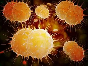
Background imageMicrobiology Collection: Microscopic view of Neisseria gonorrhoeae

Microscopic view of Neisseria gonorrhoeae. Neisseria gonorrhoeae typically infects the mucous membranes causing infections such as urethritis, cervicitis, salpingitis, pelvic inflammatory disease

Background imageMicrobiology Collection: Cluster of HIV virus

Cluster of HIV virus. HIV is the human immunodeficiency virus that can lead to acquired immune deficiency syndrom, or AIDS

Background imageMicrobiology Collection: Microscopic view of yellow fever virus

Microscopic view of yellow fever virus. Yellow fever is an acute viral disease

Background imageMicrobiology Collection: Microscopic view of Giardiasis

Microscopic view of Giardiasis, an infectious disease caused by a unicellular parasite known as Giardia lamblia

Background imageMicrobiology Collection: Microscopic view of bacterial pneumonia

Microscopic view of bacterial pneumonia. Bacterial pneumonia is a type of pneumonia caused by bacterial infection. Pneumonia can be generally defined as inflammation of the lung parenchyma

Background imageMicrobiology Collection: Microscopic view of DNA

Microscopic view of DNA

Background imageMicrobiology Collection: Microscopic view of Streptococcus pneumoniae

Microscopic view of Streptococcus pneumoniae, also known as pneumococcus. Pneumococcus is a Gram-positive coccus shaped pathogenic bacteria which causes many types of pneumococcal infections in

Background imageMicrobiology Collection: Different types of acne, non-inflammatory and inflammatory

Different types of acne, non-inflammatory and inflammatory

Background imageMicrobiology Collection: Conceptual image of mitochondria

Conceptual image of mitochondria

Background imageMicrobiology Collection: Microscopic view of Stachybotrys chartarum

Microscopic view of Stachybotrys chartarum, a black mold

Background imageMicrobiology Collection: Microscopic view of a bipolar neuron

Microscopic view of a bipolar neuron. A bipolar cell is a type of neuron which has two extensions. Bipolar cells are specialized sensory neurons for the transmission of special senses

Background imageMicrobiology Collection: Microscopic view of protozoa

Microscopic view of protozoa. Leishmaniasis and dysentery are examples of diseases involving protozoa, often transmitted to humans by the bite of a parasite-carrying insect

Background imageMicrobiology Collection: Anatomy of foot fungus with microscopic close-up

Anatomy of foot fungus with microscopic close-up

Background imageMicrobiology Collection: Conceptual image of rabies virus

Conceptual image of rabies virus

Background imageMicrobiology Collection: Sputnik virophage particle, artwork

Sputnik virophage particle, artwork
Sputnik virophage particle, computer artwork

Background imageMicrobiology Collection: HIV infected macrophage, SEM C018 / 8598

HIV infected macrophage, SEM C018 / 8598
HIV infected macrophage. Coloured ion-abrasion scanning electron micrograph (IA-SEM) of a macrophage white blood cell infected with human immunodeficiency virus (HIV, red)

Background imageMicrobiology Collection: Cell membrane lipid bilayer, artwork F007 / 1477

Cell membrane lipid bilayer, artwork F007 / 1477
Phospholipid bilayer. Computer artwork of the phospholipid bilayer that forms the membrane around all living cells. The cell membrane is made of phospholipid molecules

Background imageMicrobiology Collection: Foot-and-mouth disease virus F006 / 9556

Foot-and-mouth disease virus F006 / 9556
Foot-and-mouth disease virus. Molecular model of the foot-and-mouth disease (FMD) virus (Aphtae epizooticae) protein coat (capsid)

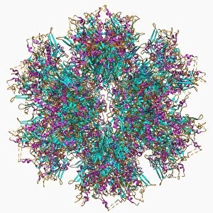
Background imageMicrobiology Collection: Adenovirus penton base protein F006 / 9542

Adenovirus penton base protein F006 / 9542
Adenovirus penton base protein, molecular model. This protein molecule is a subunit called a penton, forming the vertices of the capsid of this adenovirus

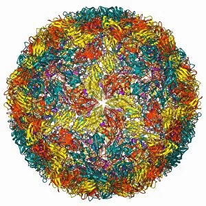
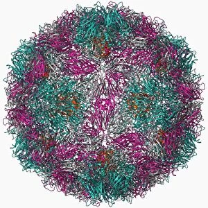
Background imageMicrobiology Collection: Rhinovirus 16 capsid, molecular model F006 / 9431

Rhinovirus 16 capsid, molecular model F006 / 9431
Rhinovirus 16 capsid, molecular model. This is human rhinovirus 16. The rhinovirus infects the upper respiratory tract and is the cause of the common cold. It is spread by coughs and sneezes

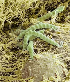
Background imageMicrobiology Collection: Helicobacter pylori bacteria, SEM C016 / 9136

Helicobacter pylori bacteria, SEM C016 / 9136
Helicobacter pylori bacteria. Coloured scanning electron micrograph (SEM) of Helicobacter pylori bacteria (green) in the stomach. H

Background imageMicrobiology Collection: Coastal wildlife, artwork C016 / 7191

Coastal wildlife, artwork C016 / 7191
Coastal wildlife. Computer artwork showing the varying forms of wildlife found at different depths on a gently sloping Mediterranean coast, with an inset showing sand-dwelling life (lower right)

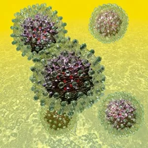
Background imageMicrobiology Collection: Hepatitis B virus particles, artwork C016 / 9097

Hepatitis B virus particles, artwork C016 / 9097
Hepatitis B virus (HBV) particles, computer artwork. Each particle consists of an outer lipid envelope (green) that surrounds a protein nucleopcapsid (purple)


All Professionally Made to Order for Quick Shipping

Microbiology, the fascinating world of tiny organisms and their impact on our lives. From the resilient Tardigrade or Water Bear that can survive extreme conditions to HeLa cells, a remarkable line of immortal human cells used in countless scientific studies. In Picture No. 11675590, we witness a mesmerizing light micrograph C017 / 8299 showcasing the intricate details of these microscopic wonders. The Neutrophil engulfing MRSA in SEM C018 / 8596 reminds us of the constant battle between our immune system and harmful bacteria. The beauty continues with another light micrograph C017 / 8298 capturing HeLa cells in all their glory. These cells have revolutionized medical research and paved the way for numerous breakthroughs. Yoghurt bacteria play a crucial role in transforming milk into this beloved creamy treat, highlighting how microbiology impacts even our daily diet. Stem cell research offers hope for regenerative medicine as we explore their incredible potential to repair and replace damaged tissues. Water bear once again graces us with its presence in light micrograph C016 / 8581, showcasing its unique anatomy under the microscope lens. We pay homage to Louis Pasteur, an influential figure who laid the foundation for modern microbiology through his groundbreaking discoveries and advancements in vaccination techniques. A glimpse into the invisible world reveals a microscopic view of human respiratory syncytial virus - a reminder that not all microbes are friendly companions. Embryo development captured just 24-36 hours after fertilization showcases nature's miracle unfolding before our eyes - each step meticulously orchestrated by molecular processes at work. And finally, artwork F008 /3245 portrays flu virus particles with artistic flair while reminding us of their ability to cause widespread illness if left unchecked. Microbiology unravels mysteries hidden from plain sight; it is both awe-inspiring and humbling as we delve deeper into this intricate realm where life's tiniest inhabitants shape our world.