
Anatomical Board Collection
The fascinating world of anatomical boards takes us on a journey through the intricate details of surgical interventions
All Professionally Made to Order for Quick Shipping
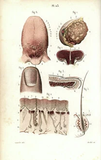
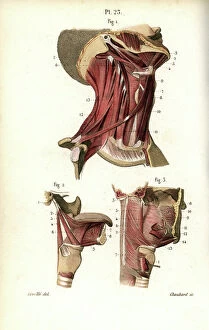
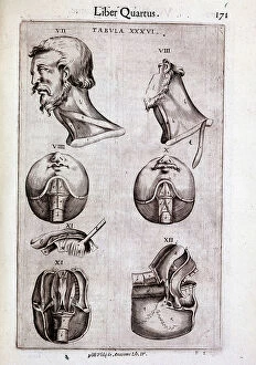
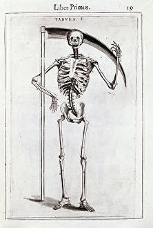
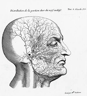
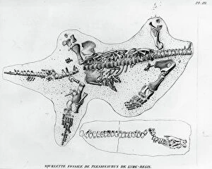
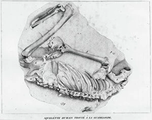
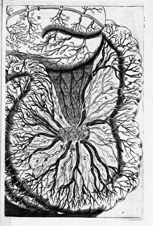
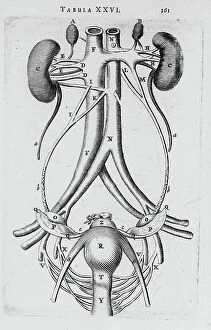
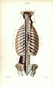
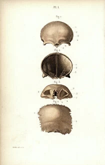
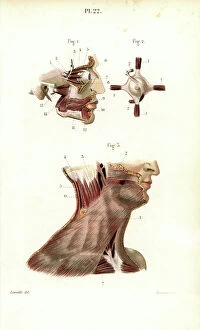
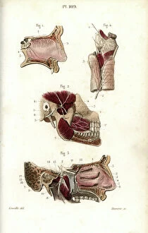
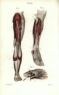
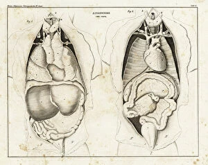
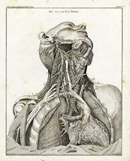
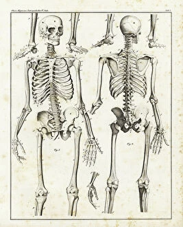
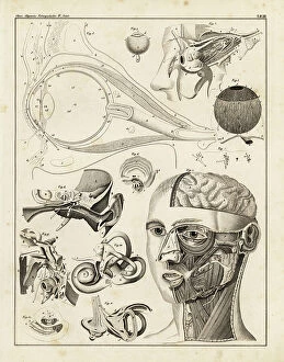
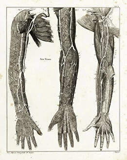
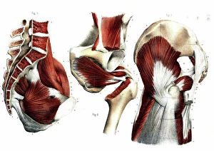
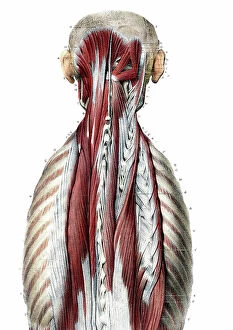
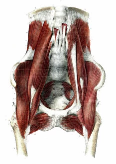
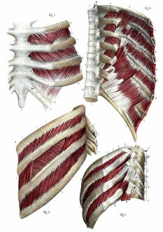
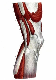
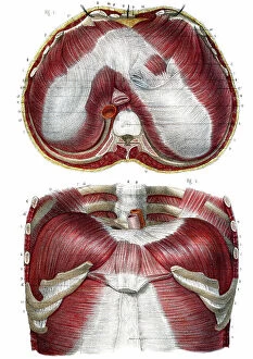
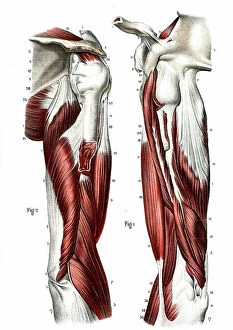
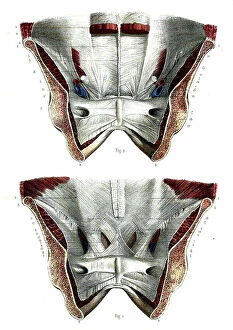
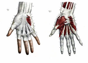
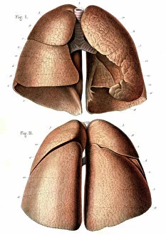
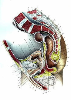
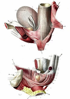
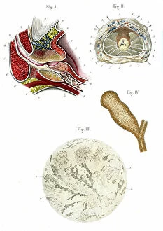
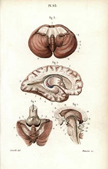
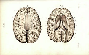
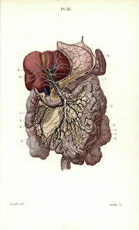
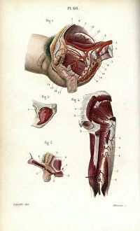
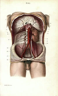
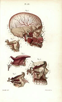
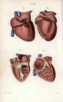
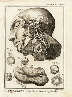
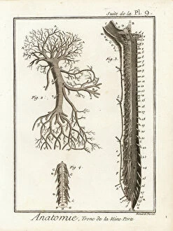
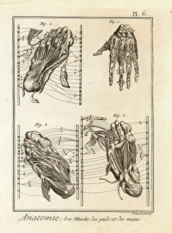
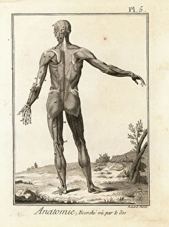
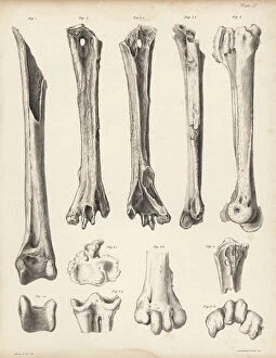
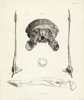
The fascinating world of anatomical boards takes us on a journey through the intricate details of surgical interventions, providing invaluable knowledge about the human body. From Walter Hermann Ryff's engraving depicting a comprehensive anatomical description of all parts of the human body, including the brain, to Dr. Rehm's Anatomical folding sheet in the New Practical Encyclopedie of Medicine and Hygiene from 1922 showcasing a naked man without apparent sex next to his skeleton - these boards offer an insight into our complex anatomy. Delving deeper into gender-specific anatomy, we encounter anatomical figures displaying male and female peels with open bellies, revealing detailed views of kidneys, bladder, and genitals. These miniature illustrations found in ancient manuscripts like "Surgery" shed light on medical practices throughout history. Moving forward in time to the late 19th century, scientific boards present cutting-edge research such as normal uterus dissections or female bodies cut at different stages of pregnancy. These visual aids were instrumental in advancing medical knowledge during that era. Not limited to just surgery-related topics, anatomical boards also explore various systems within our bodies. For instance, spinal cord localization is depicted beautifully in Gui de Pavia's "Liber notabilium Philippi septimi francorum regis, " while another section focuses on localizing male organs. Furthermore, specific attention is given to reproductive systems; one board highlights the male reproductive system with labels describing different parts such as balanos (the fommite) and Mr. Trunk (vein cava). This level of detail showcases how far our understanding has come over centuries. Anatomy isn't solely confined to surgical purposes; it has also been utilized for artistic endeavors. Engravings like those found in "Abstract Anatomy" or Giulio Casserio's "Tabulae Anatomicae" serve as references for painters and sculptors alike by providing accurate depictions of the digestive tract.