Fraud Blocker Skip to main content

Anatomical Board Collection (#2)

The fascinating world of anatomical boards takes us on a journey through the intricate details of surgical interventions

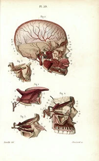
Background imageAnatomical Board Collection: The circulatory system of the brain. Lithograph of Choubard, based on a drawing by Leveille

The circulatory system of the brain. Lithograph of Choubard, based on a drawing by Leveille
FLO4672884 The circulatory system of the brain. Lithograph of Choubard, based on a drawing by Leveille, in Petity Atlas complet d'Anatomie descriptive du Corps Humain, by Dr

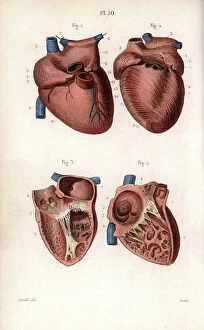
Background imageAnatomical Board Collection: Sections of the heart. Lithograph of Corbie, based on a drawing by Leveille

Sections of the heart. Lithograph of Corbie, based on a drawing by Leveille
FLO4672832 Sections of the heart. Lithograph of Corbie, based on a drawing by Leveille, in Petity Atlas complet d'Anatomie descriptive du Corps Humain, by Dr

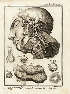
Background imageAnatomical Board Collection: Arteries in the face. Copperplate engraving by Robert Benard after an illustration by Albrecht von

Arteries in the face. Copperplate engraving by Robert Benard after an illustration by Albrecht von Haller from Denis
FLO4611624 Arteries in the face. Copperplate engraving by Robert Benard after an illustration by Albrecht von Haller from Denis Diderot's Encyclopedia, Pellet, Geneva

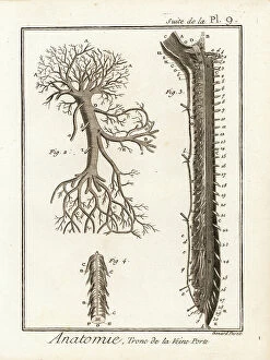
Background imageAnatomical Board Collection: Trunk of the hepatic portal vein. Copperplate engraving by Robert Benard from Denis Diderot's

Trunk of the hepatic portal vein. Copperplate engraving by Robert Benard from Denis Diderot's Encyclopedia, Pellet
FLO4611607 Trunk of the hepatic portal vein. Copperplate engraving by Robert Benard from Denis Diderot's Encyclopedia, Pellet, Geneva, 1779.; (add.info.: Trunk of the hepatic portal vein)

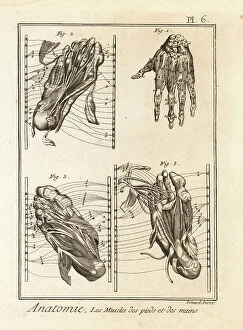
Background imageAnatomical Board Collection: Dissection of the muscles in the hand and foot. Copperplate engraving by Robert Benard from Denis

Dissection of the muscles in the hand and foot. Copperplate engraving by Robert Benard from Denis Diderot's
FLO4611571 Dissection of the muscles in the hand and foot. Copperplate engraving by Robert Benard from Denis Diderot's Encyclopedia, Pellet, Geneva

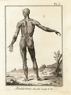
Background imageAnatomical Board Collection: Flayed human body revealing muscle system and tendons seen from the rear

Flayed human body revealing muscle system and tendons seen from the rear. Copperplate engraving by Robert Benard after
FLO4611562 Flayed human body revealing muscle system and tendons seen from the rear. Copperplate engraving by Robert Benard after an illustration by Bernhard Albinus from Denis Diderot's

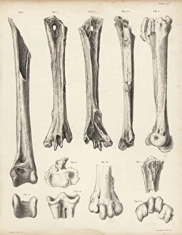
Background imageAnatomical Board Collection: Tibia and metatarsus of the extinct Rodrigues solitaire, Pezophaps solitaria

Tibia and metatarsus of the extinct Rodrigues solitaire, Pezophaps solitaria
FLO4612197 Tibia and metatarsus of the extinct Rodrigues solitaire, Pezophaps solitaria, in the Parisian Collection and Andersonian Collection

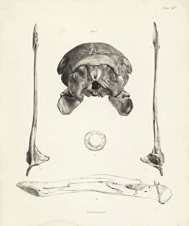
Background imageAnatomical Board Collection: Back view of skull, upper and lower view of lower jaw, inner view of jaw

Back view of skull, upper and lower view of lower jaw, inner view of jaw, and circle of sclerotic bones in the dodo
FLO4612158 Back view of skull, upper and lower view of lower jaw, inner view of jaw, and circle of sclerotic bones in the dodo

Background imageAnatomical Board Collection: Anatomy of the human nose, mouth, tongue, jaw and teeth, showing the senses of taste

Anatomy of the human nose, mouth, tongue, jaw and teeth, showing the senses of taste, smell and touch
FLO4684860 Anatomy of the human nose, mouth, tongue, jaw and teeth, showing the senses of taste, smell and touch. Lithograph from Lorenz Oken's Universal Natural History

Background imageAnatomical Board Collection: Cranium 1-4 and part of sternum 5, 6 of the extinct Rodrigues solitaire

Cranium 1-4 and part of sternum 5, 6 of the extinct Rodrigues solitaire. Lithograph by Joseph Dinkel after Werner
FLO4612182 Cranium 1-4 and part of sternum 5, 6 of the extinct Rodrigues solitaire. Lithograph by Joseph Dinkel after Werner from Hugh Edwin Strickland

Background imageAnatomical Board Collection: Skulls of tooth-billed pigeon, Didunculus strigirostris, critically endangered 1, 7, 8, 9, 10 dodo

Skulls of tooth-billed pigeon, Didunculus strigirostris, critically endangered 1, 7, 8, 9, 10 dodo
FLO4612171 Skulls of tooth-billed pigeon, Didunculus strigirostris, critically endangered 1, 7, 8, 9, 10 dodo, Raphus cucullatus 2, yellow-footed green pigeon, Treron chlorigaster 3, 6

Background imageAnatomical Board Collection: Base of the skull, human teeth, knee cap (after Clopton Havers), mucus gland, etc

Base of the skull, human teeth, knee cap (after Clopton Havers), mucus gland, etc
FLO4611538 Base of the skull, human teeth, knee cap (after Clopton Havers), mucus gland, etc. Copperplate engraving by Robert Benard from Denis Diderot's Encyclopedia, Pellet, Geneva

Background imageAnatomical Board Collection: Interior of the brain and cerebellum. Copperplate engraving by Robert Benard after an illustration

Interior of the brain and cerebellum. Copperplate engraving by Robert Benard after an illustration by Albrecht von
FLO4611652 Interior of the brain and cerebellum. Copperplate engraving by Robert Benard after an illustration by Albrecht von Haller from Denis Diderot's Encyclopedia, Pellet, Geneva

Background imageAnatomical Board Collection: Arteries in the face. Copperplate engraving by Robert Benard after an illustration by Albrecht von

Arteries in the face. Copperplate engraving by Robert Benard after an illustration by Albrecht von Haller from Denis
FLO4611618 Arteries in the face. Copperplate engraving by Robert Benard after an illustration by Albrecht von Haller from Denis Diderot's Encyclopedia, Pellet, Geneva

Background imageAnatomical Board Collection: Venal system from the vena cava in the human body. Copperplate engraving by Robert Benard after an

Venal system from the vena cava in the human body. Copperplate engraving by Robert Benard after an illustration by
FLO4611599 Venal system from the vena cava in the human body. Copperplate engraving by Robert Benard after an illustration by James Drake from Denis Diderot's Encyclopedia, Pellet, Geneva

Background imageAnatomical Board Collection: Details of arteries, veins, lymph acorns, etc. Copperplate engraving by Robert Benard after

Details of arteries, veins, lymph acorns, etc. Copperplate engraving by Robert Benard after from Denis Diderot's
FLO4611594 Details of arteries, veins, lymph acorns, etc. Copperplate engraving by Robert Benard after from Denis Diderot's Encyclopedia, Pellet, Geneva, 1779.; (add.info.: Details of arteries)

Background imageAnatomical Board Collection: Upper and lower views of the skull of a dodo. Lithograph from Hugh Edwin Strickland

Upper and lower views of the skull of a dodo. Lithograph from Hugh Edwin Strickland
FLO4612153 Upper and lower views of the skull of a dodo. Lithograph from Hugh Edwin Strickland and Alexander Gordon Melville's The Dodo and its Kindred, London, Reeve, Benham and Reeve

Background imageAnatomical Board Collection: Tooth billed pigeon, Didunculus strigirostris 1, 2 (critically endangered), Bruce's green pigeon

Tooth billed pigeon, Didunculus strigirostris 1, 2 (critically endangered), Bruce's green pigeon
FLO4612139 Tooth billed pigeon, Didunculus strigirostris 1, 2 (critically endangered), Bruce's green pigeon, Treron waalia (Treron abyssinica 3, 5) and Levaillant's dove

Background imageAnatomical Board Collection: Bladder anatomy 1866 illustrations showing the internal (top) and external (bottom)

Bladder anatomy 1866 illustrations showing the internal (top) and external (bottom)
JAB4119242 Bladder anatomy 1866 illustrations showing the internal (top) and external (bottom) anatomy of the male urinary bladder, showing its muscular coating (brown)

Background imageAnatomical Board Collection: Voice box and thyroid 1866 illustration showing the anatomy of the larynx (voice box)

Voice box and thyroid 1866 illustration showing the anatomy of the larynx (voice box) and thyroid (brown)
JAB4119213 Voice box and thyroid 1866 illustration showing the anatomy of the larynx (voice box) and thyroid (brown, lower centre) Shown here are the hyoid bone (A), thyroid cartilage (B)

Background imageAnatomical Board Collection: Chest muscles - 1866 illustration showing the structure of the muscles (brown) and tendons (white)

Chest muscles - 1866 illustration showing the structure of the muscles (brown) and tendons (white) of the human chest
JAB4119479 Chest muscles - 1866 illustration showing the structure of the muscles (brown) and tendons (white) of the human chest

Background imageAnatomical Board Collection: Leg and foot muscles - 1866 anatomical illustration showing the muscles (brown) and tendons (white)

Leg and foot muscles - 1866 anatomical illustration showing the muscles (brown) and tendons (white)
JAB4119476 Leg and foot muscles - 1866 anatomical illustration showing the muscles (brown) and tendons (white) of the right foot (left)

Background imageAnatomical Board Collection: Shoulder muscles - 1866 illustration of the muscles (brown) and tendons (white)

Shoulder muscles - 1866 illustration of the muscles (brown) and tendons (white)
JAB4119462 Shoulder muscles - 1866 illustration of the muscles (brown) and tendons (white) associated with the human collarbone (clavicle, top) and shoulder blade (scapula)

Background imageAnatomical Board Collection: Chest and shoulder muscles - 1866 anatomical illustration showing the structure of the muscles

Chest and shoulder muscles - 1866 anatomical illustration showing the structure of the muscles (brown)
JAB4119433 Chest and shoulder muscles - 1866 anatomical illustration showing the structure of the muscles (brown) and tendons (white)

Background imageAnatomical Board Collection: Hand muscles - 1866 anatomical illustration showing the muscles (brown) and tendons (white)

Hand muscles - 1866 anatomical illustration showing the muscles (brown) and tendons (white) of the human hand
JAB4119426 Hand muscles - 1866 anatomical illustration showing the muscles (brown) and tendons (white) of the human hand, with insets (left)

Background imageAnatomical Board Collection: Rib cage anatomy - 1866 illustration showing the bones of the human rib cage

Rib cage anatomy - 1866 illustration showing the bones of the human rib cage, including the ribs (bottom)
JAB4119411 Rib cage anatomy - 1866 illustration showing the bones of the human rib cage, including the ribs (bottom), spine (centre-left) and sternum (breast bone, centre and upper right) Le Thorax

Background imageAnatomical Board Collection: Pelvis anatomy - 1866 illustration showing the anatomy of the male (top) and female (bottom)

Pelvis anatomy - 1866 illustration showing the anatomy of the male (top) and female (bottom) pelvis - The pelvis
JAB4119404 Pelvis anatomy - 1866 illustration showing the anatomy of the male (top) and female (bottom) pelvis - The pelvis, anatomy, osteology - Fig 1: female pelvis Fig 2: male pelvis, Musculotion

Background imageAnatomical Board Collection: Hip muscles - 1866 illustration showing the structure of the muscles (brown) and tendons (white)

Hip muscles - 1866 illustration showing the structure of the muscles (brown) and tendons (white)
JAB4119401 Hip muscles - 1866 illustration showing the structure of the muscles (brown) and tendons (white) of the upper right leg and hip

Background imageAnatomical Board Collection: Arm muscles - 1866 illustration showing the structure of the muscles (brown) and tendons (white)

Arm muscles - 1866 illustration showing the structure of the muscles (brown) and tendons (white)
JAB4119391 Arm muscles - 1866 illustration showing the structure of the muscles (brown) and tendons (white) of the human arm and top of the hand (bottom left and right)

Background imageAnatomical Board Collection: Abdominal muscles - 1866 illustration showing the structure of the muscles (brown)

Abdominal muscles - 1866 illustration showing the structure of the muscles (brown) and tendons (white)
JAB4119387 Abdominal muscles - 1866 illustration showing the structure of the muscles (brown) and tendons (white) found in the rear of the human trunk - L'abdomen - Anatomy - Region anterieure de

Background imageAnatomical Board Collection: Leg muscles - 1866 illustration showing the structure of the muscles (brown) and tendons (white)

Leg muscles - 1866 illustration showing the structure of the muscles (brown) and tendons (white)
JAB4119363 Leg muscles - 1866 illustration showing the structure of the muscles (brown) and tendons (white) of the rear of the lower leg - Anatomy du dos de la leg - Fig 1 et 2

Background imageAnatomical Board Collection: Head muscles. Lithograph of Lebrun, based on a drawing by Leveille

Head muscles. Lithograph of Lebrun, based on a drawing by Leveille, in Petity Atlas complet d'Anatomie descriptive
FLO4672607 Head muscles. Lithograph of Lebrun, based on a drawing by Leveille, in Petity Atlas complet d'Anatomie descriptive du Corps Humain, by Dr

Background imageAnatomical Board Collection: Hip joint and thigh bone. Lithograph of Corbie, in Petity Atlas complet d'Anatomie descriptive du

Hip joint and thigh bone. Lithograph of Corbie, in Petity Atlas complet d'Anatomie descriptive du Corps Humain
FLO4672595 Hip joint and thigh bone. Lithograph of Corbie, in Petity Atlas complet d'Anatomie descriptive du Corps Humain, by Dr

Background imageAnatomical Board Collection: Sympathic nervous system in the thoracic, lumbar and sacral plexus. Lithograph by Davesne

Sympathic nervous system in the thoracic, lumbar and sacral plexus. Lithograph by Davesne
FLO4672529 Sympathic nervous system in the thoracic, lumbar and sacral plexus. Lithograph by Davesne, based on a drawing by Leveille

Background imageAnatomical Board Collection: Sympathic nervous system in the cardiac plexus. Lithograph by Davesne

Sympathic nervous system in the cardiac plexus. Lithograph by Davesne, based on a drawing by Leveille
FLO4672521 Sympathic nervous system in the cardiac plexus. Lithograph by Davesne, based on a drawing by Leveille, in Petiure Atlas complet d'Anatomie descriptive du Corps Humain, by Dr

Background imageAnatomical Board Collection: Nervous system of the back. Lithograph of Corbie, based on a drawing by Leveille

Nervous system of the back. Lithograph of Corbie, based on a drawing by Leveille
FLO4672457 Nervous system of the back. Lithograph of Corbie, based on a drawing by Leveille, in Petity Atlas complet d'Anatomie descriptive du Corps Humain, by Dr

Background imageAnatomical Board Collection: Facial and auditory nerves. Lithograph by Davesne, based on a drawing by Leveille

Facial and auditory nerves. Lithograph by Davesne, based on a drawing by Leveille
FLO4672442 Facial and auditory nerves. Lithograph by Davesne, based on a drawing by Leveille, in Petite Atlas complet d'Anatomie descriptive du Corps Humain, by Dr

Background imageAnatomical Board Collection: Intercostal nerves. Lithograph of Corbie, based on a drawing by Leveille

Intercostal nerves. Lithograph of Corbie, based on a drawing by Leveille, in Petity Atlas complet d'Anatomie
FLO4672415 Intercostal nerves. Lithograph of Corbie, based on a drawing by Leveille, in Petity Atlas complet d'Anatomie descriptive du Corps Humain, by Dr

Background imageAnatomical Board Collection: Fetus head helped through the birth canal with forceps. Copperplate engraving by Andrew Bell after

Fetus head helped through the birth canal with forceps. Copperplate engraving by Andrew Bell after an illustration by
FLO4695943 Fetus head helped through the birth canal with forceps. Copperplate engraving by Andrew Bell after an illustration by Jan van Rymsdyk from William Smellie's A Set of Anatomical Tables

Background imageAnatomical Board Collection: Le Coeur, extract from 'The anatomy of the human body' 1797, by Charles Bell (1774-1824)

Le Coeur, extract from "The anatomy of the human body" 1797, by Charles Bell (1774-1824)
XEE5028095 Le Coeur, extract from "The anatomy of the human body" 1797, by Charles Bell (1774-1824) - Biblioteca nazionale braidense, Milano; (add.info.: Le Coeur)

Background imageAnatomical Board Collection: Anatomy: representation of the uterus by Garioponto (970 - 1050), Italian doctor of the Middle Ages

Anatomy: representation of the uterus by Garioponto (970 - 1050), Italian doctor of the Middle Ages
XEE5029574 Anatomy: representation of the uterus by Garioponto (970 - 1050), Italian doctor of the Middle Ages, Biblioteca Civica Bertoliana

Background imageAnatomical Board Collection: Metoposcopiae liber, The metoposcopy of 1658, by (Cardanus Hieronymus) (Jerome Cardane)

Metoposcopiae liber, The metoposcopy of 1658, by (Cardanus Hieronymus) (Jerome Cardane) Gerolamo Cardano (1501-1576)
XEE5028461 Metoposcopiae liber, The metoposcopy of 1658, by (Cardanus Hieronymus) (Jerome Cardane) Gerolamo Cardano (1501-1576) Italian doctor, mathematician

Background imageAnatomical Board Collection: External female parts of generation, labia, clitoris, perineum, and anus

External female parts of generation, labia, clitoris, perineum, and anus. Copperplate engraving by Andrew Bell after an
FLO4696243 External female parts of generation, labia, clitoris, perineum, and anus. Copperplate engraving by Andrew Bell after an illustration by Jan van Rymsdyk from William Smellie's A Set of

Background imageAnatomical Board Collection: Deep nervous system of the arm. Lithograph by Davesne, based on a drawing by Leveille

Deep nervous system of the arm. Lithograph by Davesne, based on a drawing by Leveille
FLO4673176 Deep nervous system of the arm. Lithograph by Davesne, based on a drawing by Leveille, in Petity Atlas complet d'Anatomie descriptive du Corps Humain, by Dr

Background imageAnatomical Board Collection: Brain, pons, spinal bulb, pineal gland etc... Lithograph by Davesne, based on a drawing by Leveille

Brain, pons, spinal bulb, pineal gland etc... Lithograph by Davesne, based on a drawing by Leveille
FLO4673157 Brain, pons, spinal bulb, pineal gland etc... Lithograph by Davesne, based on a drawing by Leveille, in Petity Atlas complet d'Anatomie descriptive du Corps Humain, by Dr

Background imageAnatomical Board Collection: Brachial plexus. Lithograph by Davesne, based on a drawing by Leveille

Brachial plexus. Lithograph by Davesne, based on a drawing by Leveille, in Petity Atlas complet d'Anatomie
FLO4673154 Brachial plexus. Lithograph by Davesne, based on a drawing by Leveille, in Petity Atlas complet d'Anatomie descriptive du Corps Humain, by Dr

Background imageAnatomical Board Collection: Section through the brain. Lithograph by Davesne, based on a drawing by Leveille

Section through the brain. Lithograph by Davesne, based on a drawing by Leveille
FLO4673112 Section through the brain. Lithograph by Davesne, based on a drawing by Leveille, in Petity Atlas complet d'Anatomie descriptive du Corps Humain, by Dr

Background imageAnatomical Board Collection: Different sections of the skull. Lithograph of Corbie, based on a drawing by Leveille

Different sections of the skull. Lithograph of Corbie, based on a drawing by Leveille
FLO4673087 Different sections of the skull. Lithograph of Corbie, based on a drawing by Leveille, in Petity Atlas complet d'Anatomie descriptive du Corps Humain, by Dr


All Professionally Made to Order for Quick Shipping

The fascinating world of anatomical boards takes us on a journey through the intricate details of surgical interventions, providing invaluable knowledge about the human body. From Walter Hermann Ryff's engraving depicting a comprehensive anatomical description of all parts of the human body, including the brain, to Dr. Rehm's Anatomical folding sheet in the New Practical Encyclopedie of Medicine and Hygiene from 1922 showcasing a naked man without apparent sex next to his skeleton - these boards offer an insight into our complex anatomy. Delving deeper into gender-specific anatomy, we encounter anatomical figures displaying male and female peels with open bellies, revealing detailed views of kidneys, bladder, and genitals. These miniature illustrations found in ancient manuscripts like "Surgery" shed light on medical practices throughout history. Moving forward in time to the late 19th century, scientific boards present cutting-edge research such as normal uterus dissections or female bodies cut at different stages of pregnancy. These visual aids were instrumental in advancing medical knowledge during that era. Not limited to just surgery-related topics, anatomical boards also explore various systems within our bodies. For instance, spinal cord localization is depicted beautifully in Gui de Pavia's "Liber notabilium Philippi septimi francorum regis, " while another section focuses on localizing male organs. Furthermore, specific attention is given to reproductive systems; one board highlights the male reproductive system with labels describing different parts such as balanos (the fommite) and Mr. Trunk (vein cava). This level of detail showcases how far our understanding has come over centuries. Anatomy isn't solely confined to surgical purposes; it has also been utilized for artistic endeavors. Engravings like those found in "Abstract Anatomy" or Giulio Casserio's "Tabulae Anatomicae" serve as references for painters and sculptors alike by providing accurate depictions of the digestive tract.