Fraud Blocker Skip to main content

Anatomical Board Collection (#4)

The fascinating world of anatomical boards takes us on a journey through the intricate details of surgical interventions

Background imageAnatomical Board Collection: Lung anatomy - 1866 illustration showing the external structure of the left (top) and right (bottom)

Lung anatomy - 1866 illustration showing the external structure of the left (top) and right (bottom)
JAB4119370 Lung anatomy - 1866 illustration showing the external structure of the left (top) and right (bottom) lungs - Lung - Descriptive anatomy - Fig 1: right lung, outer face, Fig 2: left lung

Background imageAnatomical Board Collection: Thoracic spine anatomy - 1866 anatomical illustration of the thoracic spine

Thoracic spine anatomy - 1866 anatomical illustration of the thoracic spine
JAB4119368 Thoracic spine anatomy - 1866 anatomical illustration of the thoracic spine, showing the individual vertebrae - Vertebral column - Dorsal vertebrae, osteology, locomotion

Background imageAnatomical Board Collection: Human throat 1866 cutaway illustration showing the anatomy of the throat LE LARYNX

Human throat 1866 cutaway illustration showing the anatomy of the throat LE LARYNX
JAB4119196 Human throat 1866 cutaway illustration showing the anatomy of the throat LE LARYNX, posterieure face - Fig 1: Pharynx and esophagus, Fig 2: Opening of the larynx 5 sup-glottic part

Background imageAnatomical Board Collection: Voice box anatomy 1866 anatomical illustration showing the muscles (brown)

Voice box anatomy 1866 anatomical illustration showing the muscles (brown) and external (Figs 1 and 2)
JAB4119181 Voice box anatomy 1866 anatomical illustration showing the muscles (brown) and external (Figs 1 and 2) and internal structure (Fig 3) of the larynx (voice box) Muscles du larynx - Fig 1

Background imageAnatomical Board Collection: Diaphragm 1866

Diaphragm 1866
JAB4119165 Diaphragm 1866; (add.info.: Diaphragm 1866 illustration of a section through the abdomen and chest, showing the diaphragm (arched)

Background imageAnatomical Board Collection: Menstrual cycle anatomy 1866

Menstrual cycle anatomy 1866
JAB4119155 Menstrual cycle anatomy 1866; (add.info.: Menstrual cycle anatomy 1866 illustrations showing the internal anatomy of a human ovary containing Graafian (ovarian) follicles (Fig 1)

Background imageAnatomical Board Collection: Male pelvic blood supply 1866 illustration showing the major veins (blue) and arteries (red)

Male pelvic blood supply 1866 illustration showing the major veins (blue) and arteries (red)
JAB4119143 Male pelvic blood supply 1866 illustration showing the major veins (blue) and arteries (red) of the male pelvis These include the inferior vena cava, common iliac vein

Background imageAnatomical Board Collection: Anatomy of the human arterial system in the legs. Handcoloured lithograph by St

Anatomy of the human arterial system in the legs. Handcoloured lithograph by St
FLO4684812 Anatomy of the human arterial system in the legs. Handcoloured lithograph by St. Schillinger from Lorenz Oken's Universal Natural History, Allgemeine Naturgeschichte fur alle Stande

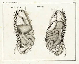
Background imageAnatomical Board Collection: Anatomy of human intestines from the side. Lithograph from Lorenz Oken's Universal Natural History

Anatomy of human intestines from the side. Lithograph from Lorenz Oken's Universal Natural History
FLO4684807 Anatomy of human intestines from the side. Lithograph from Lorenz Oken's Universal Natural History, Allgemeine Naturgeschichte fur alle Stande, Stuttgart

Background imageAnatomical Board Collection: Brain sections. Lithograph by Davesne, based on a drawing by Leveille

Brain sections. Lithograph by Davesne, based on a drawing by Leveille, in Petity Atlas complet d'Anatomie
FLO4673204 Brain sections. Lithograph by Davesne, based on a drawing by Leveille, in Petity Atlas complet d'Anatomie descriptive du Corps Humain, by Dr

Background imageAnatomical Board Collection: Vertebral column, ribs, vertebrae and sternum. Lithograph of Corbie

Vertebral column, ribs, vertebrae and sternum. Lithograph of Corbie, in Petity Atlas complet d'Anatomie descriptive
FLO4673029 Vertebral column, ribs, vertebrae and sternum. Lithograph of Corbie, in Petity Atlas complet d'Anatomie descriptive du Corps Humain, by Dr

Background imageAnatomical Board Collection: Section through the abdomen. Lithograph of Lebrun, based on a drawing by Leveille

Section through the abdomen. Lithograph of Lebrun, based on a drawing by Leveille
FLO4672840 Section through the abdomen. Lithograph of Lebrun, based on a drawing by Leveille, in Petity Atlas complet d'Anatomie descriptive du Corps Humain, by Dr

Background imageAnatomical Board Collection: Aponevrosis (fibrous membrane that covers and supports the muscles) of the torso

Aponevrosis (fibrous membrane that covers and supports the muscles) of the torso, cross section and frontal view
FLO4672748 Aponevrosis (fibrous membrane that covers and supports the muscles) of the torso, cross section and frontal view

Background imageAnatomical Board Collection: Aponevrosis of the neck, shoulder and eye. Lithograph of Corbie, based on a drawing by Leveille

Aponevrosis of the neck, shoulder and eye. Lithograph of Corbie, based on a drawing by Leveille
FLO4672728 Aponevrosis of the neck, shoulder and eye. Lithograph of Corbie, based on a drawing by Leveille, in Petiure Atlas complet d'Anatomie descriptive du Corps Humain, by Dr

Background imageAnatomical Board Collection: Muscles and tendons of the leg and foot. Lithograph of Lebrun, based on a drawing by Leveille

Muscles and tendons of the leg and foot. Lithograph of Lebrun, based on a drawing by Leveille
FLO4672719 Muscles and tendons of the leg and foot. Lithograph of Lebrun, based on a drawing by Leveille, in Petity Atlas complet d'Anatomie descriptive du Corps Humain, by Dr

Background imageAnatomical Board Collection: Thigh and knee muscles and tendons. Lithograph of Lebrun, based on a drawing by Leveille

Thigh and knee muscles and tendons. Lithograph of Lebrun, based on a drawing by Leveille
FLO4672703 Thigh and knee muscles and tendons. Lithograph of Lebrun, based on a drawing by Leveille, in Petity Atlas complet d'Anatomie descriptive du Corps Humain, by Dr

Background imageAnatomical Board Collection: Thigh muscles and tendons. Lithograph of Lebrun, based on a drawing by Leveille

Thigh muscles and tendons. Lithograph of Lebrun, based on a drawing by Leveille
FLO4672702 Thigh muscles and tendons. Lithograph of Lebrun, based on a drawing by Leveille, in Petity Atlas complet d'Anatomie descriptive du Corps Humain, by Dr

Background imageAnatomical Board Collection: Muscles and tendons of the forearm and wrist. Lithograph by Davesne, based on a drawing by Leveille

Muscles and tendons of the forearm and wrist. Lithograph by Davesne, based on a drawing by Leveille
FLO4672691 Muscles and tendons of the forearm and wrist. Lithograph by Davesne, based on a drawing by Leveille, in Petity Atlas complet d'Anatomie descriptive du Corps Humain, by Dr

Background imageAnatomical Board Collection: Muscles of the forearm and wrist. Lithograph by Davesne, based on a drawing by Leveille

Muscles of the forearm and wrist. Lithograph by Davesne, based on a drawing by Leveille
FLO4672685 Muscles of the forearm and wrist. Lithograph by Davesne, based on a drawing by Leveille, in Petity Atlas complet d'Anatomie descriptive du Corps Humain, by Dr

Background imageAnatomical Board Collection: Shoulder and arm muscles. Lithograph of Anneshower, based on a drawing by Leveille

Shoulder and arm muscles. Lithograph of Anneshower, based on a drawing by Leveille
FLO4672670 Shoulder and arm muscles. Lithograph of Anneshower, based on a drawing by Leveille, in Petiure Atlas complet d'Anatomie descriptive du Corps Humain, by Dr

Background imageAnatomical Board Collection: Internal muscles of the back. Lithograph by Davesne, based on a drawing by Leveille

Internal muscles of the back. Lithograph by Davesne, based on a drawing by Leveille
FLO4672652 Internal muscles of the back. Lithograph by Davesne, based on a drawing by Leveille, in Petity Atlas complet d'Anatomie descriptive du Corps Humain, by Dr

Background imageAnatomical Board Collection: Torso muscles (male). Lithograph of anneshower, based on a drawing by Leveille

Torso muscles (male). Lithograph of anneshower, based on a drawing by Leveille
FLO4672627 Torso muscles (male). Lithograph of anneshower, based on a drawing by Leveille, in Petiter Atlas complet d'Anatomie descriptive du Corps Humain, by Dr

Background imageAnatomical Board Collection: Shoulder and elbow joints. Lithograph of Corbie, in Petity Atlas complet d'Anatomie descriptive du

Shoulder and elbow joints. Lithograph of Corbie, in Petity Atlas complet d'Anatomie descriptive du Corps Humain
FLO4672545 Shoulder and elbow joints. Lithograph of Corbie, in Petity Atlas complet d'Anatomie descriptive du Corps Humain, by Dr

Background imageAnatomical Board Collection: Hand bone: carps, metacarps and phalanges and leg: femur and shin. Lithograph of Corbie

Hand bone: carps, metacarps and phalanges and leg: femur and shin. Lithograph of Corbie
FLO4672512 Hand bone: carps, metacarps and phalanges and leg: femur and shin. Lithograph of Corbie, in Petity Atlas complet d'Anatomie descriptive du Corps Humain, by Dr

Background imageAnatomical Board Collection: Cervical plexus. Lithograph of Corbie, based on a drawing by Leveille

Cervical plexus. Lithograph of Corbie, based on a drawing by Leveille, in Petity Atlas complet d'Anatomie
FLO4673165 Cervical plexus. Lithograph of Corbie, based on a drawing by Leveille, in Petity Atlas complet d'Anatomie descriptive du Corps Humain, by Dr

Background imageAnatomical Board Collection: Veins of the legs and foot. Lithograph of Corbie, based on a drawing by Leveille

Veins of the legs and foot. Lithograph of Corbie, based on a drawing by Leveille
FLO4673005 Veins of the legs and foot. Lithograph of Corbie, based on a drawing by Leveille, in Petity Atlas complet d'Anatomie descriptive du Corps Humain, by Dr

Background imageAnatomical Board Collection: Axis and vertebrae. Lithograph of Corbie, in Petity Atlas complet d'Anatomie descriptive du Corps

Axis and vertebrae. Lithograph of Corbie, in Petity Atlas complet d'Anatomie descriptive du Corps Humain, by Dr
FLO4672967 Axis and vertebrae. Lithograph of Corbie, in Petity Atlas complet d'Anatomie descriptive du Corps Humain, by Dr

Background imageAnatomical Board Collection: Circulatory system of the intestines. Lithograph of Anneshower, based on a drawing by Leveille

Circulatory system of the intestines. Lithograph of Anneshower, based on a drawing by Leveille
FLO4672922 Circulatory system of the intestines. Lithograph of Anneshower, based on a drawing by Leveille, in Petity Atlas complet d'Anatomie descriptive du Corps Humain, by Dr

Background imageAnatomical Board Collection: Back and torso muscles. Lithograph by Davesne, based on a drawing by Leveille

Back and torso muscles. Lithograph by Davesne, based on a drawing by Leveille
FLO4672626 Back and torso muscles. Lithograph by Davesne, based on a drawing by Leveille, in Petity Atlas complet d'Anatomie descriptive du Corps Humain, by Dr

Background imageAnatomical Board Collection: Pelvic bone, front and back views. Lithograph of Corbie, in Petity Atlas complet d'Anatomie

Pelvic bone, front and back views. Lithograph of Corbie, in Petity Atlas complet d'Anatomie descriptive du Corps
FLO4672563 Pelvic bone, front and back views. Lithograph of Corbie, in Petity Atlas complet d'Anatomie descriptive du Corps Humain, by Dr

Background imageAnatomical Board Collection: Vertebral column, spine and neck. Lithograph of Corbie, in Petity Atlas complet d'Anatomie

Vertebral column, spine and neck. Lithograph of Corbie, in Petity Atlas complet d'Anatomie descriptive du Corps
FLO4672546 Vertebral column, spine and neck. Lithograph of Corbie, in Petity Atlas complet d'Anatomie descriptive du Corps Humain, by Dr

Background imageAnatomical Board Collection: Glossopharyngeal nerve. Lithograph by Davesne, based on a drawing by Leveille

Glossopharyngeal nerve. Lithograph by Davesne, based on a drawing by Leveille
FLO4672515 Glossopharyngeal nerve. Lithograph by Davesne, based on a drawing by Leveille, in Petity Atlas complet d'Anatomie descriptive du Corps Humain, by Dr

Background imageAnatomical Board Collection: Nerves of the skull and eye. Lithograph by Davesne, based on a drawing by Leveille

Nerves of the skull and eye. Lithograph by Davesne, based on a drawing by Leveille
FLO4672497 Nerves of the skull and eye. Lithograph by Davesne, based on a drawing by Leveille, in Petity Atlas complet d'Anatomie descriptive du Corps Humain, by Dr

Background imageAnatomical Board Collection: Ophthalmic and maxillary nerves of the fifth cranial nerve. Lithograph by Davesne

Ophthalmic and maxillary nerves of the fifth cranial nerve. Lithograph by Davesne, based on a drawing by Leveille
FLO4672496 Ophthalmic and maxillary nerves of the fifth cranial nerve. Lithograph by Davesne, based on a drawing by Leveille, in Petity Atlas complet d'Anatomie descriptive du Corps Humain

Background imageAnatomical Board Collection: Lumbar plexus and crural nerve. Lithograph of Corbie, based on a drawing by Leveille

Lumbar plexus and crural nerve. Lithograph of Corbie, based on a drawing by Leveille
FLO4672455 Lumbar plexus and crural nerve. Lithograph of Corbie, based on a drawing by Leveille, in Petity Atlas complet d'Anatomie descriptive du Corps Humain, by Dr

Background imageAnatomical Board Collection: Shoulder and arm bone: scapula, humerus, cubitus and radius. Lithograph of Corbie

Shoulder and arm bone: scapula, humerus, cubitus and radius. Lithograph of Corbie
FLO4672433 Shoulder and arm bone: scapula, humerus, cubitus and radius. Lithograph of Corbie, in Petity Atlas complet d'Anatomie descriptive du Corps Humain, by Dr

Background imageAnatomical Board Collection: Asthma. Engraving from ' Les Grands Ales et les Grands Remedes' by Jules Rengade, 1889

Asthma. Engraving from " Les Grands Ales et les Grands Remedes" by Jules Rengade, 1889
PCT4274721 Asthma. Engraving from " Les Grands Ales et les Grands Remedes" by Jules Rengade, 1889.; (add.info.: Asthma)

Background imageAnatomical Board Collection: Newborn male and female reproductive organs 1866 cutaway computer artwork comparing the anatomy of

Newborn male and female reproductive organs 1866 cutaway computer artwork comparing the anatomy of the reproductive
JAB4119239 Newborn male and female reproductive organs 1866 cutaway computer artwork comparing the anatomy of the reproductive system of a newborn boy (left) and girl (right)

Background imageAnatomical Board Collection: Uterus blood supply 1866 illustration showing the major blood vessels supplying the uterus (womb)

Uterus blood supply 1866 illustration showing the major blood vessels supplying the uterus (womb)
JAB4119232 Uterus blood supply 1866 illustration showing the major blood vessels supplying the uterus (womb) in a female human Arteres du bassin chez la femme - Arteres de uterus

Background imageAnatomical Board Collection: Lung anatomy 1866 illustrations showing the external anatomy of the human lungs (red) Fig 1

Lung anatomy 1866 illustrations showing the external anatomy of the human lungs (red) Fig 1: from below
JAB4119217 Lung anatomy 1866 illustrations showing the external anatomy of the human lungs (red) Fig 1: from below, showing the arteries (centre) Fig 2: frontal view, showing the bronchi (centre)

Background imageAnatomical Board Collection: Upper body muscles 1866 illustration showing the muscle structure of the chest

Upper body muscles 1866 illustration showing the muscle structure of the chest
JAB4119145 Upper body muscles 1866 illustration showing the muscle structure of the chest and neck Laterale region of the trunk 2 - occipital bone, external auditory duct, styloid apophysis

Background imageAnatomical Board Collection: Larynx, trachee artere and bronchi - The normal life and health of J. Rengade.Drawing A

Larynx, trachee artere and bronchi - The normal life and health of J. Rengade.Drawing A. Demarle 1881
JAB4120247 Larynx, trachee artere and bronchi - The normal life and health of J. Rengade.Drawing A. Demarle 1881; (add.info.: Larynx, trachee artere and bronchi - The normal life and health of J)

Background imageAnatomical Board Collection: Nursed children (The normal life & health of J. Rengade) - Top: breast-fed child

Nursed children (The normal life & health of J. Rengade) - Top: breast-fed child, left-fed child fed with porridge
JAB4120168 Nursed children (The normal life & health of J. Rengade) - Top: breast-fed child, left-fed child fed with porridge. Bottom right: Ricky child.Drawing by A

Background imageAnatomical Board Collection: Human thigh muscles - 1866 illustration showing the structure of the muscles (brown)

Human thigh muscles - 1866 illustration showing the structure of the muscles (brown) and tendons (white)
JAB4119468 Human thigh muscles - 1866 illustration showing the structure of the muscles (brown) and tendons (white) of the rear of the human thigh and hip, showing the sacrum (white, left)

Background imageAnatomical Board Collection: Leg muscles - 1866 illustration showing the structure of the muscles (brown) and tendons (white)

Leg muscles - 1866 illustration showing the structure of the muscles (brown) and tendons (white)
JAB4119456 Leg muscles - 1866 illustration showing the structure of the muscles (brown) and tendons (white) of the rear of the lower leg and ankle (bottom)

Background imageAnatomical Board Collection: Leg muscles - 1866 illustration showing the structure of the muscles (brown) and tendons (white)

Leg muscles - 1866 illustration showing the structure of the muscles (brown) and tendons (white)
JAB4119442 Leg muscles - 1866 illustration showing the structure of the muscles (brown) and tendons (white) of the rear of the upper leg - Region femoral posterieure - Fig 1: symphysis of the pubis

Background imageAnatomical Board Collection: Leg and knee muscles - 1866 anatomical illustration showing the muscles (brown) and tendons (white)

Leg and knee muscles - 1866 anatomical illustration showing the muscles (brown) and tendons (white) of the human leg
JAB4119421 Leg and knee muscles - 1866 anatomical illustration showing the muscles (brown) and tendons (white) of the human leg

Background imageAnatomical Board Collection: Abdominal muscles - 1866 illustration showing the structure of the muscles (brown)

Abdominal muscles - 1866 illustration showing the structure of the muscles (brown) and tendons (white)
JAB4119415 Abdominal muscles - 1866 illustration showing the structure of the muscles (brown) and tendons (white) found in the human abdomen (centre)


All Professionally Made to Order for Quick Shipping

The fascinating world of anatomical boards takes us on a journey through the intricate details of surgical interventions, providing invaluable knowledge about the human body. From Walter Hermann Ryff's engraving depicting a comprehensive anatomical description of all parts of the human body, including the brain, to Dr. Rehm's Anatomical folding sheet in the New Practical Encyclopedie of Medicine and Hygiene from 1922 showcasing a naked man without apparent sex next to his skeleton - these boards offer an insight into our complex anatomy. Delving deeper into gender-specific anatomy, we encounter anatomical figures displaying male and female peels with open bellies, revealing detailed views of kidneys, bladder, and genitals. These miniature illustrations found in ancient manuscripts like "Surgery" shed light on medical practices throughout history. Moving forward in time to the late 19th century, scientific boards present cutting-edge research such as normal uterus dissections or female bodies cut at different stages of pregnancy. These visual aids were instrumental in advancing medical knowledge during that era. Not limited to just surgery-related topics, anatomical boards also explore various systems within our bodies. For instance, spinal cord localization is depicted beautifully in Gui de Pavia's "Liber notabilium Philippi septimi francorum regis, " while another section focuses on localizing male organs. Furthermore, specific attention is given to reproductive systems; one board highlights the male reproductive system with labels describing different parts such as balanos (the fommite) and Mr. Trunk (vein cava). This level of detail showcases how far our understanding has come over centuries. Anatomy isn't solely confined to surgical purposes; it has also been utilized for artistic endeavors. Engravings like those found in "Abstract Anatomy" or Giulio Casserio's "Tabulae Anatomicae" serve as references for painters and sculptors alike by providing accurate depictions of the digestive tract.