Metal Print > Arts > Artists > E > Thomas Ender
Metal Print : Synapse nerve junction, TEM

![]()
Metal Prints from Science Photo Library
Synapse nerve junction, TEM
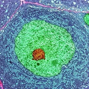
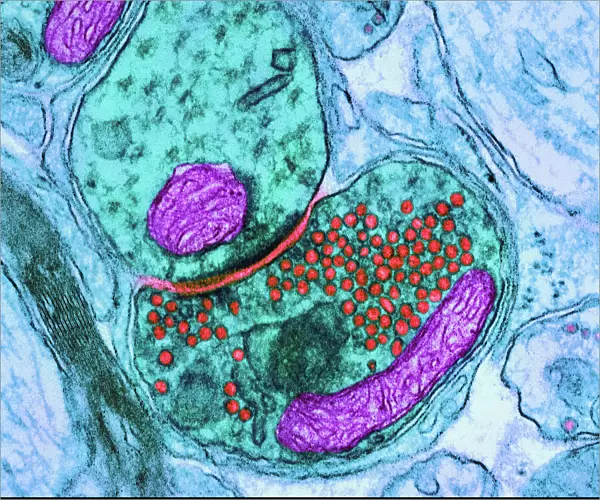
Synapse. Coloured transmission electron micrograph (TEM) of a synapse, a junction between two nerve cells, in the brain. At a synapse an electrical signal is transmitted from one cell to the next in only one direction. The nerve cells are green, with the pre-synaptic cell at lower right and the post-synaptic cell at upper left. Mitochondria, supplying the cells with energy, are purple. When an electrical signal reaches a synapse it triggers the release of neurotransmitter chemicals from vesicles (red) at the end of the presynaptic cell. The neurotransmitters cross a microscopic gap and bind to receptors on the post- synaptic cell. Magnification: 50, 000x when printed 10 centimetres across
Science Photo Library features Science and Medical images including photos and illustrations
Media ID 6449159
© THOMAS DEERINCK, NCMIR/SCIENCE PHOTO LIBRARY
Axon Central Cerebral Grey Matter Histological Histology Junction Message Micrograph Nerve Cell Nervous Neural Neuron Neurone Neurones Neurons Physiological Physiology Relay Relaying Signal Signals Synapse Synaptic System Transmission Electron Transmission Electron Microscope Transmitting Vesicle Vesicles Brain Cells False Coloured Nervous System Neurological Neurology
20"x24" (61x51cm) Metal Print
Discover the intricacies of the natural world with our Media Storehouse Metal Prints featuring this stunning Transmission Electron Micrograph (TEM) of a Synapse nerve junction from Science Photo Library. Witness the complex interplay between two nerve cells as an electrical signal is transmitted, bringing science to life in vibrant colors and sharp detail on high-quality metal. Each print is meticulously crafted to bring out the true essence of this colossal discovery, making it a captivating addition to any scientific or home decor.
Made with durable metal and luxurious printing techniques, our metal photo prints go beyond traditional canvases, adding a cool, modern touch to your space. Wall mount on back. Eco-friendly 100% post-consumer recycled ChromaLuxe aluminum surface. The thickness of the print is 0.045". Featuring a Scratch-resistant surface and Rounded corners. Backing hangers are attached to the back of the print and float the print 1/2-inch off the wall when hung, the choice of hanger may vary depending on size and International orders will come with Float Mount hangers only. Finished with a brilliant white high gloss surface for unsurpassed detail and vibrance. Printed using Dye-Sublimation and for best care we recommend a non-ammonia glass cleaner, water, or isopropyl (rubbing) alcohol to prevent harming the print surface. We recommend using a clean, lint-free cloth to wipe off the print. The ultra-hard surface is scratch-resistant, waterproof and weatherproof. Avoid direct sunlight exposure.
Made with durable metal and luxurious printing techniques, metal prints bring images to life and add a modern touch to any space
Estimated Image Size (if not cropped) is 60.9cm x 50.8cm (24" x 20")
Estimated Product Size is 61.5cm x 51.4cm (24.2" x 20.2")
These are individually made so all sizes are approximate
Artwork printed orientated as per the preview above, with landscape (horizontal) orientation to match the source image.
FEATURES IN THESE COLLECTIONS
> Arts
> Artists
> C
> Thomas Cross
> Arts
> Artists
> E
> Thomas Ender
EDITORS COMMENTS
This print showcases the intricate beauty of a synapse nerve junction, captured using a transmission electron microscope (TEM). The image reveals the remarkable complexity and functionality of this vital component in our brain's neural network. Colored to enhance visual clarity, it depicts two nerve cells connected at a synapse, where electrical signals are transmitted unidirectionally. The green-hued nerve cells stand out against the background, with the pre-synaptic cell positioned at the lower right and the post-synaptic cell at upper left. Providing energy to these cells are mitochondria depicted in striking purple. When an electrical signal reaches this synapse, it triggers vesicles (depicted in red) within the presynaptic cell to release neurotransmitter chemicals. These neurotransmitters traverse a microscopic gap before binding to receptors on the post-synaptic cell. This crucial process allows for efficient communication between neurons and is fundamental to our nervous system's functioning. With a magnification level of 50,000x when printed at 10 centimeters across, this image offers us an awe-inspiring glimpse into one of nature's most intricate mechanisms. As we explore this photograph further, we gain insight into neurology and physiology while marveling at its anatomical details. It serves as a reminder that even within our own bodies lie wonders waiting to be discovered through scientific exploration and understanding.
MADE IN THE USA
Safe Shipping with 30 Day Money Back Guarantee
FREE PERSONALISATION*
We are proud to offer a range of customisation features including Personalised Captions, Color Filters and Picture Zoom Tools
SECURE PAYMENTS
We happily accept a wide range of payment options so you can pay for the things you need in the way that is most convenient for you
* Options may vary by product and licensing agreement. Zoomed Pictures can be adjusted in the Cart.