
Cell Division Collection
"Unlocking the Mysteries of Life: Exploring Cell Division Through Micrographs and Illustrations" Witness the intricate dance of life as cells divide through mitosis


All Professionally Made to Order for Quick Shipping
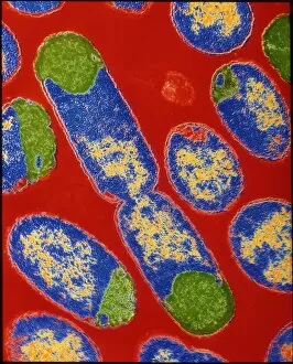
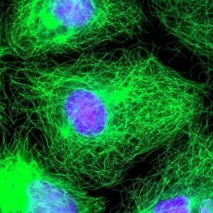
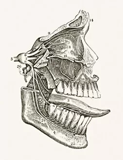
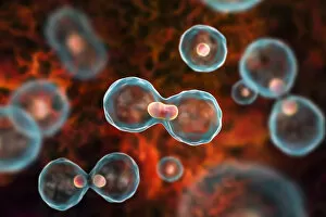
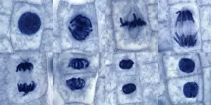
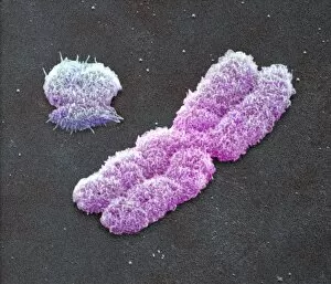
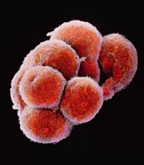
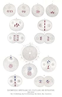
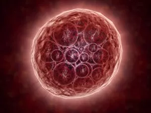
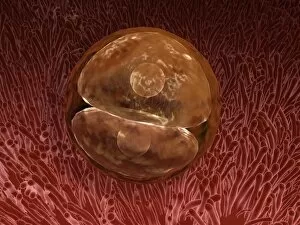
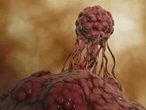
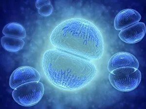
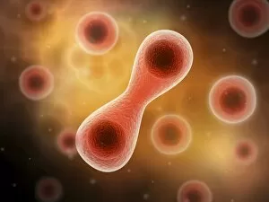
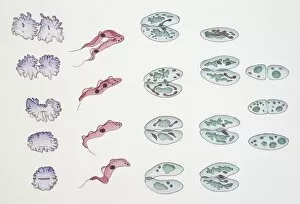
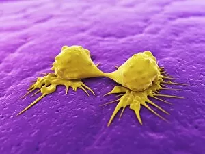
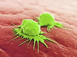
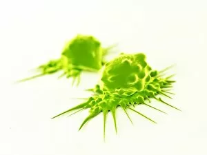
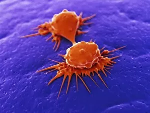
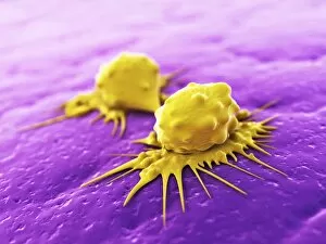
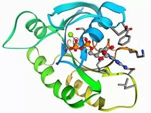
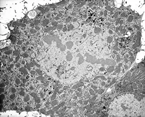
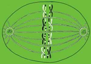
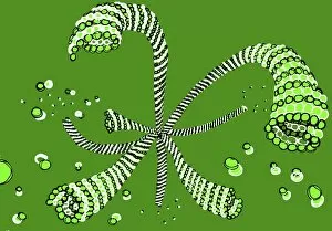
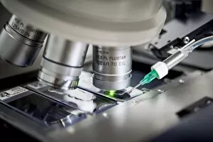
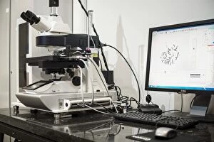
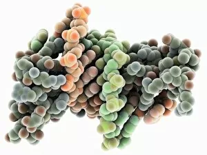
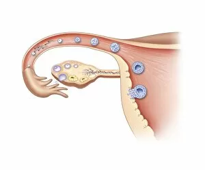
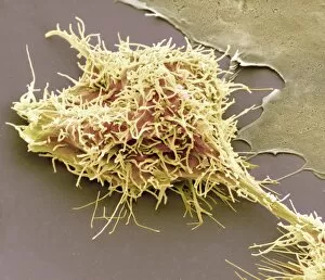
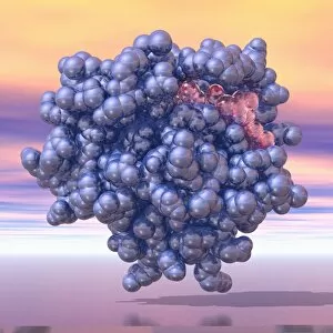
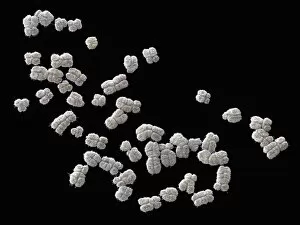
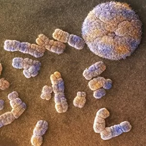
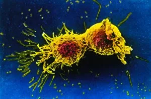
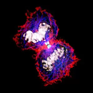
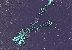
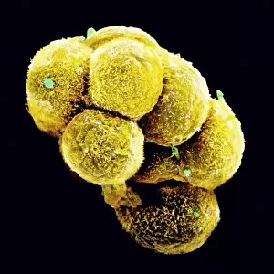
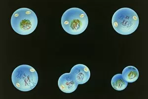
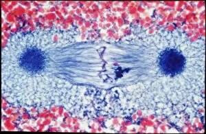
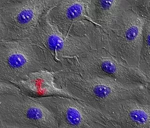
"Unlocking the Mysteries of Life: Exploring Cell Division Through Micrographs and Illustrations" Witness the intricate dance of life as cells divide through mitosis, captured in a mesmerizing light micrograph. Delve into the microscopic world of E. Coli bacteria, where a constant process, revealed in a fluorescent micrograph. Marvel at the astonishing progress of embryo development just 24-36 hours after fertilization, depicted in another captivating fluorescent micrograph. Explore the inner workings of an E. Coli bacterium under high magnification with a TEM image that showcases its dividing nature. Immerse yourself in the vibrant false-color TEM image that unveils the beauty within an E. coli bacterium during division. Observe with awe as an E. coli bacterium undergoes division, revealing its remarkable ability to multiply and thrive. Journey into human biology as SEM captures striking images of chromosomes during cell division – a crucial process for growth and repair. Uncover the intricacies of cellular reproduction through SEM imagery that highlights various stages across different organisms. Step back in time with a fascinating 19th-century medical illustration showcasing facial nerves involved in complex processes like cell division. Explore nuclear division through an illuminating illustration that unravels how genetic material is precisely distributed during this vital cellular event. In this captivating collection, we embark on an enlightening journey through various scientific techniques and illustrations to unravel one of life's most fundamental processes – cell division.