Fraud Blocker Skip to main content

Cell Division Collection (#2)

"Unlocking the Mysteries of Life: Exploring Cell Division Through Micrographs and Illustrations" Witness the intricate dance of life as cells divide through mitosis

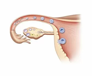
Background imageCell Division Collection: Ovulation and fertilisation, artwork

Ovulation and fertilisation, artwork
Ovulation and fertilisation. Computer artwork of the female reproductive system, showing the development of an ovum (egg, small circle) inside an ovary (oval)

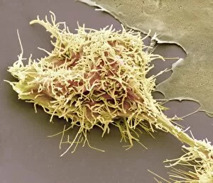
Background imageCell Division Collection: Dividing brain cancer cells, SEM C014 / 0354

Dividing brain cancer cells, SEM C014 / 0354
Dividing brain cancer cells. Coloured scanning electron micrograph (SEM) of a cancerous astrocyte brain cell that has just undergone cytokinesis (cell division)

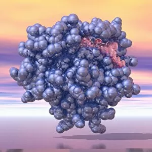
Background imageCell Division Collection: HRas enzyme molecule C014 / 0010

HRas enzyme molecule C014 / 0010
HRas enzyme molecule. Molecular model showing the structure of a molecule of the enzyme GTPase HRas, also known as transforming protein p21

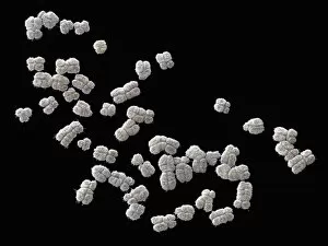
Background imageCell Division Collection: Set of human chromosomes, SEM

Set of human chromosomes, SEM
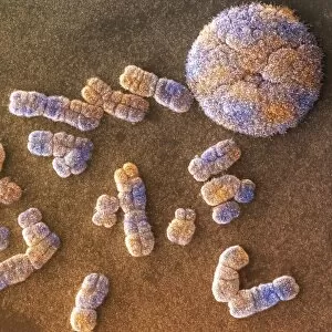
Set of human chromosomes, coloured scanning election micrograph (SEM). Chromosomes are a packaged form of the genetic material deoxyribonucleic acid (DNA)

Background imageCell Division Collection: Human chromosomes and nucleus, SEM

Human chromosomes and nucleus, SEM
Human chromosomes and nucleus, coloured scanning electron micrograph (SEM). Chromosomes are a packaged form of the genetic material DNA (deoxyribonucleic acid)

Background imageCell Division Collection: Scanning electron micrograph of cell division

Scanning electron micrograph of cell division
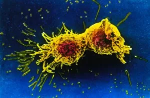
Cell division. Coloured scanning electron micrograph of two human cells pulling apart in the final stage or telophase of cell division

Background imageCell Division Collection: Cytokinesis

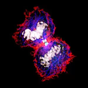
Cytokinesis. Fluorescent micrograph of an animal cell during cytokinesis (cell division). Cytokinesis occurs after nuclear division (mitosis), which produces two daughter nuclei

Background imageCell Division Collection: Lampbrush chromosomes, TEM

Lampbrush chromosomes, TEM
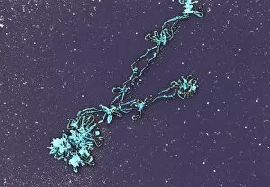
Lampbrush chromosomes. Coloured transmission electron micrograph (TEM) of lampbrush chromosomes (LBCs). A chromosome consists of proteins and DNA (deoxyribonucleic acid)

Background imageCell Division Collection: Coloured SEM of an embryo at the stage of morula

Coloured SEM of an embryo at the stage of morula
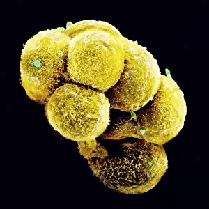
Embryo development. Coloured scanning electron micrograph of an embryo at the early stage known as the morula. The egg reaches this phase about 4 days after fertilisation after a series of mitotic

Background imageCell Division Collection: Illustration of the first stage of Meiosis

Illustration of the first stage of Meiosis
Illustration of the first stage of meiosis (gametogenesis), a two-stage cell division process exclusive to production of male and female gametes in sexual reproduction

Background imageCell Division Collection: LM of metaphase mitosis in a triton (sea slug) egg

LM of metaphase mitosis in a triton (sea slug) egg
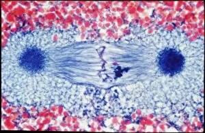
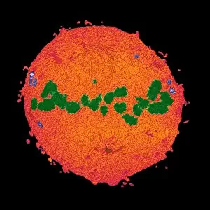
Metaphase cell division. Light micrograph of cell division (mitosis) in a triton (naked sea slug/ nudibranch) egg. This is the metaphase stage in mitosis

Background imageCell Division Collection: Dividing cell

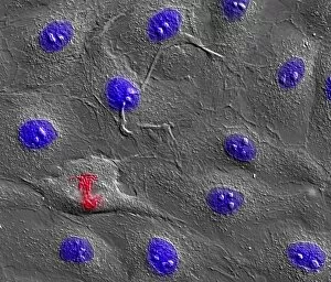
Dividing cell. Differential interference contrast (DIC) light micrograph of a cell (lower left) in the metaphase stage of mitosis (cell division). The cells nuclei are stained with fluorescent dye

Background imageCell Division Collection: Mitotic cell division, TEM

Mitotic cell division, TEM
Mitotic cell division. Coloured transmission electron micrograph (TEM) of a cell during the metaphase stage of mitosis (cell division)

Background imageCell Division Collection: Mitosis, TEM

Mitosis, TEM
Mitosis, coloured transmission electron micrograph (TEM). Longitudinal section through a human T cell in metaphase. During mitosis two daughter nuclei are formed from one parent nucleus

Background imageCell Division Collection: Cell mitosis

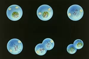
Cell mitosis. Confocal fluorescence light micrograph composite showing 6 stages of mitotic cell division. At far left, the cell has completed the first stage of cell division known as interphase in

Background imageCell Division Collection: Cell division

Cell division. Fluorescent micrograph of an animal cell during cytokinesis (cell division). Cytokinesis occurs after nuclear division (mitosis)

Background imageCell Division Collection: Lymph node, light micrograph

Lymph node, light micrograph
Lymph node. Coloured light micrograph of a section through a lymph node. A lymph node filters pathogens from lymph fluid, a watery liquid that surrounds the tissues of the body

Background imageCell Division Collection: Stem cells and God

Stem cells and God. Conceptual computer artwork showing enlarged cells in a petri dish, with a representation of God from Michelangelos painting The Creation of Adam

Background imageCell Division Collection: Stem cell on spoon

Stem cell on spoon. Conceptual computer artwork showing an enlarged stem cell on a spoon. A stem cell is an undifferentiated cell that can produce other types of cell when it divides

Background imageCell Division Collection: HaCaT culture cell, light micrograph

HaCaT culture cell, light micrograph
HaCaT cell. Immunofluorescence light micrograph of a HaCaT cell dividing into two. The nucleus, which contains the cells genetic information, is purple

Background imageCell Division Collection: Mitosis, artwork

Mitosis, artwork

Background imageCell Division Collection: Enterococcus faecalis, bacteria, artwork

Enterococcus faecalis, bacteria, artwork
Enterococcus faecalis. Computer artwork of Enterococcus faecalis bacteria (previously known as Streptococcus faecalis). The bacterium is in the process of dividing through binary fission. E

Background imageCell Division Collection: Protocell proliferation, artwork

Protocell proliferation, artwork
Protocell proliferation. Image 5 of 5. Artwork showing a protocell (artificial cell) dividing to produce two daughter cells

Background imageCell Division Collection: Stem cells division, computer artwork

Stem cells division, computer artwork
Computer artwork depicting division of stem cells. A stem cell is an undifferentiated call that can divide indefinitely in culture

Background imageCell Division Collection: Lily flower bud, light micrograph

Lily flower bud, light micrograph
Lily flower bud. Light micrograph of a section through a young flower bud from a lily (Lilium sp.), showing pollen cells (circles) dividing within the anthers (inner ring)

Background imageCell Division Collection: Human chromosome 16, SEM

Human chromosome 16, SEM
Human chromosome 16, coloured scanning electron micrograph (SEM). Chromosomes are a packaged form of the genetic material DNA (deoxyribonucleic acid)

Background imageCell Division Collection: Human chromosome 10, SEM

Human chromosome 10, SEM
Human chromosome 10, coloured scanning electron micrograph (SEM). Chromosomes are a packaged form of the genetic material DNA (deoxyribonucleic acid)

Background imageCell Division Collection: Cytokinesis, artwork

Cytokinesis, artwork
Cytokinesis. Artwork showing the stage of cell division that involves the splitting of the cell cytoplasm between two daughter cells

Background imageCell Division Collection: Cell division, artwork

Cell division, artwork
Cell division. Computer artwork of an animal cell undergoing mitosis (nuclear division) and cytokinesis (cell division) to form two identical daughter cells (bottom)

Background imageCell Division Collection: Cytokinesis, diagram

Cytokinesis, diagram
Cytokinesis. Diagram showing the stage of cell division that involves the splitting of the cell cytoplasm between two daughter cells

Background imageCell Division Collection: Plant cell wall, diagram

Plant cell wall, diagram

Background imageCell Division Collection: Cell division, conceptual artwork

Cell division, conceptual artwork
cell division

Background imageCell Division Collection: Bacteria circadian clock protein molecule

Bacteria circadian clock protein molecule. Computer model showing the molecular structure of the KaiC protein found in cyanobacteria (blue-green algae)

Background imageCell Division Collection: Chromosomes, artwork

Chromosomes, artwork
Chromosomes, computer artwork. Chromosomes are a packaged form of the genetic material DNA (deoxyribonucleic acid). The DNA condenses into chromosomes during cell replication for ease of division


All Professionally Made to Order for Quick Shipping

"Unlocking the Mysteries of Life: Exploring Cell Division Through Micrographs and Illustrations" Witness the intricate dance of life as cells divide through mitosis, captured in a mesmerizing light micrograph. Delve into the microscopic world of E. Coli bacteria, where a constant process, revealed in a fluorescent micrograph. Marvel at the astonishing progress of embryo development just 24-36 hours after fertilization, depicted in another captivating fluorescent micrograph. Explore the inner workings of an E. Coli bacterium under high magnification with a TEM image that showcases its dividing nature. Immerse yourself in the vibrant false-color TEM image that unveils the beauty within an E. coli bacterium during division. Observe with awe as an E. coli bacterium undergoes division, revealing its remarkable ability to multiply and thrive. Journey into human biology as SEM captures striking images of chromosomes during cell division – a crucial process for growth and repair. Uncover the intricacies of cellular reproduction through SEM imagery that highlights various stages across different organisms. Step back in time with a fascinating 19th-century medical illustration showcasing facial nerves involved in complex processes like cell division. Explore nuclear division through an illuminating illustration that unravels how genetic material is precisely distributed during this vital cellular event. In this captivating collection, we embark on an enlightening journey through various scientific techniques and illustrations to unravel one of life's most fundamental processes – cell division.