
Thorax Collection (#4)
"Exploring the Marvels of the Thorax: From Ancient Art to Modern Medicine" The thorax, a vital region housing the heart and lungs


All Professionally Made to Order for Quick Shipping
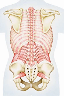
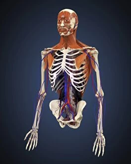
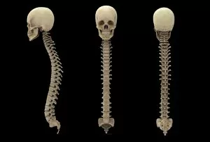
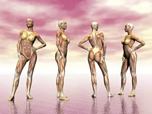
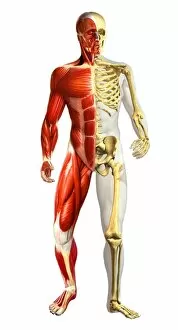
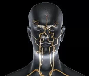
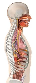
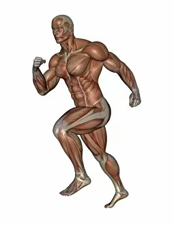
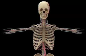
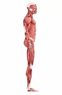
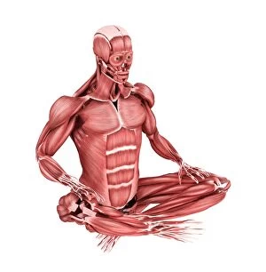
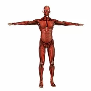
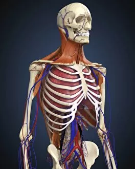
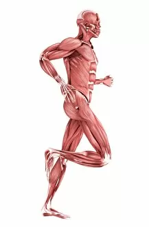
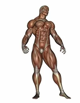
"Exploring the Marvels of the Thorax: From Ancient Art to Modern Medicine" The thorax, a vital region housing the heart and lungs, is truly a masterpiece of human anatomy. An athletic young man in shorts lies on a stone floor, his thoracic muscles glistening with sweat after an intense workout. Delicate veins and arteries form an intricate web within the cardiovascular system, ensuring oxygen-rich blood reaches every corner of our body. Behold historical artwork depicting ancient sculptures that beautifully capture the essence of the thorax's strength and grace. In 19th-century artwork, detailed depictions of neck anatomy shed light on how this crucial part connects our head to our torso. X-ray images reveal the devastating impact tuberculosis can have on delicate structures within the thorax. A honey bee viewed through a scanning electron microscope showcases its astonishingly complex internal structure within its tiny thorax. Irregular heartbeat? Consult your doctor for proper diagnosis and treatment options to keep your precious thoracic organ healthy. Julien Bougle's stunning illustration overlays colored plates onto a human body, highlighting various systems including the intricate details of the thorax. The hornet mimic hoverfly flaunts its deceptive appearance as it hovers near flowers, showcasing its remarkable resemblance to dangerous predators despite being harmless itself. Dive into microscopic wonders as we explore cross-sections revealing female mosquitoes' internal anatomy while they feed by sucking blood from our skin – nature's fascinating yet pesky creatures. A man with a naked torso sits amidst bales of hay in a horse barn; his exposed chest reminds us all of how vulnerable but resilient our thoraxes are.