
Swine Flu Collection (#2)
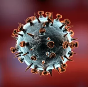
"Unveiling the Swine Flu: A Closer Look at the Influenza Virus" In this captivating image, we dive into the microscopic world of swine flu, also known as H1N1 influenza


All Professionally Made to Order for Quick Shipping
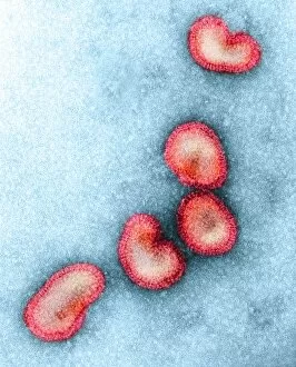
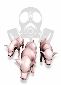
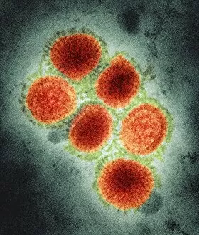
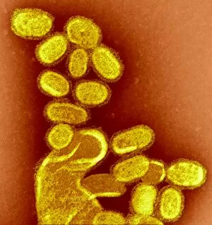
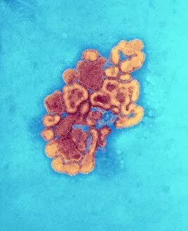
"Unveiling the Swine Flu: A Closer Look at the Influenza Virus" In this captivating image, we dive into the microscopic world of swine flu, also known as H1N1 influenza. Through a transmission electron microscope (TEM), we witness an intricate dance of influenza virus particles, revealing their unique structure and composition. The first conceptual image portrays the notorious influenza-causing flu virus in all its glory. Its spherical shape is adorned with spikes that enable it to latch onto host cells and initiate infection. This visual representation serves as a reminder of the invisible threat lurking around us during flu seasons. Moving forward, our attention shifts to a group of H5N1 viruses captured under a glassy view. These strains have garnered significant attention due to their potential for causing severe illness in both animals and humans. The delicate balance between fascination and fear arises from witnessing these tiny entities capable of wreaking havoc on our health. Returning to another conceptual image, we encounter yet again the influenza-causing flu virus - a constant reminder that vigilance against respiratory diseases remains crucial for public health worldwide. It reminds us how easily this infectious agent can spread through coughs and sneezes, making prevention measures imperative. A closer examination reveals blood cells invaded by these minuscule viral invaders – an unsettling sight indeed. Microscopic view showcases how these viruses hijack our own cells' machinery to replicate themselves rapidly, leading to symptoms such as fever, body ache, sore throat, and respiratory distress. Once more confronted with a group of H5N1 viruses viewed through glassy lenses; their presence emphasizes the urgency for continued research efforts aimed at understanding their behavior better while developing effective vaccines or antiviral treatments. Finally, we arrive at an iconic depiction of swine influenza virus itself – responsible for outbreaks among pigs but occasionally crossing over into human populations too. This snapshot encapsulates not only its distinctive appearance but also the potential danger it poses to our health.