
Skeletal System Collection (#5)
The intricate beauty of the skeletal system revealed through art and science. From skeletons to X-ray artwork, explore the fascinating world within our bones


All Professionally Made to Order for Quick Shipping
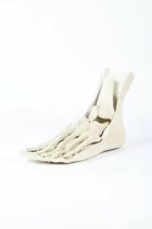
The intricate beauty of the skeletal system revealed through art and science. From skeletons to X-ray artwork, explore the fascinating world within our bones. Marvel at the complexity of a skeleton from below, depicted in stunning X-ray artwork. Delve into biomechanics with historical artwork showcasing the interplay between structure and movement. Witness the flexibility of neck vertebrae extended in an X-ray image, highlighting our body's remarkable adaptability. Gain a comprehensive understanding of the human spine with a detailed diagram from a side view. Discover the inner workings of our mouth and jaw through a cross-section diagram, unveiling their role in speech and digestion. Explore every aspect of foot anatomy - skin, veins, arteries, muscles, and bones - all working together for locomotion. Contemplate life's fragility with a conceptual image featuring a human skull alongside its spinal cord; reminding us that we are more than just flesh and blood. Uncover different angles of human skull anatomy through captivating visuals that showcase its intricate design. Stand in awe as you witness the entirety of the human skeletal system from front view – an architectural marvel supporting our bodies throughout life's journey. Dive deeper into this wondrous framework with an anterior view accompanied by labels that reveal each bone's unique identity. The skeletal system is not merely about bones; it is about strength, resilience, mobility – it is what holds us upright and allows us to move forward.