Fraud Blocker Skip to main content

Single Celled Collection (#6)

"Unveiling the Microscopic World: Captivating Single-Celled Organisms Under the SEM" Delicate and intricate

Background imageSingle Celled Collection: Evolution of life, artwork

Evolution of life, artwork
Evolution of life. Computer artwork showing the evolution of animal and plant life from single-celled organisms (bottom left) to modern humans today (right centre)

Background imageSingle Celled Collection: Paramecium protozoan, artwork

Paramecium protozoan, artwork
Paramecium protozoan. Computer artwork of a Paramecium sp. ciliate protozoan. Its single cell is covered in numerous cilia (white lines), hair-like structures that it beats for propulsion

Background imageSingle Celled Collection: Protozoan tentacle, TEM

Protozoan tentacle, TEM
Protozoan tentacle. Coloured transmission electron micrograph (TEM) of a cross-section through a tentacle of a Dendrocometes sp. protozoan showing the microtubule (purple circles) structure

Background imageSingle Celled Collection: Trichodina parasite, SEM

Trichodina parasite, SEM
Trichodina parasite. Coloured scanning electron micrograph (SEM) of a Trichodina sp. protist on a freshwater minnow. This ciliate parasite lives on the skin

Background imageSingle Celled Collection: Diatom shell, SEM

Diatom shell, SEM
Diatom shell. Coloured scanning electron micrograph (SEM) of the shell of a Biddulphia sp. diatom. The diatoms are a group of photosynthetic, single-celled algae containing about 10, 000 species

Background imageSingle Celled Collection: Algae, SEM

Algae, SEM
Algae. Coloured scanning electron micrograph (SEM) of single-celled algae (green). Magnification: x4000 when printed 10 centimetres wide

Background imageSingle Celled Collection: Euglena flagellate protozoans, SEM

Euglena flagellate protozoans, SEM
Euglena flagellate protozoans. Scanning electron micrograph (SEM) of Euglena sp. flagellate protozoans. Euglena sp. is a freshwater unicellular (single-celled)

Background imageSingle Celled Collection: Radiolarian, SEM

Radiolarian, SEM
Radiolarian. Coloured scanning electron micrograph (SEM) of the shell of a radiolarian. Radiolaria are single-celled protozoans that are found in marine plankton

Background imageSingle Celled Collection: Actinosphaerium protozoan

Actinosphaerium protozoan. Differential interference contrast micrograph of an Actinoshaerium sp. heliozoa protozoan. This single-celled freshwater organism has many pseudopodia radiating outwards

Background imageSingle Celled Collection: Euglena protozoan, light micrograph

Euglena protozoan, light micrograph
Euglena protozoan. Differential interference contrast micrograph of the flagellate protozoan Euglena spirogyra. This freshwater single-celled organism can either obtain energy from sunlight

Background imageSingle Celled Collection: Shelled amoeba, light micrograph

Shelled amoeba, light micrograph
Shelled amoeba. Differential interference contrast micrograph of the testate (shelled) amoeba Arcella sp.. The circular shell is formed form the polysaccharide chitin

Background imageSingle Celled Collection: Euglena protozoa, light micrograph

Euglena protozoa, light micrograph
Euglena protozoa. Differential interference contrast micrograph of the flagellate protozoa Euglena acus. These freshwater single-celled organisms can either obtain energy from sunlight

Background imageSingle Celled Collection: Vorticella protozoa, light micrograph

Vorticella protozoa, light micrograph
Vorticella protozoa. Differential interference contrast micrograph of a colony of Vorticella sp.. This ciliate protozoan consists of a bell-shaped head and a stalk that is anchored to the substrate

Background imageSingle Celled Collection: Radiolarian skeleton, artwork

Radiolarian skeleton, artwork
Radiolarian skeleton. Computer artwork of the skeleton (test) of a radiolarian. Radiolaria are single-celled protozoans that are found in marine plankton

Background imageSingle Celled Collection: Ciliate protozoan, SEM

Ciliate protozoan, SEM
Ciliate protozoan. Coloured scanning electron micrograph (SEM) of a Paramecium cilliate protozoan. Protozoa are a type of unicellular animal

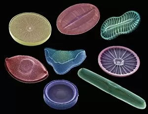
Background imageSingle Celled Collection: Selection of diatoms, SEM

Selection of diatoms, SEM
Selection of diatoms, coloured scanning electron micrograph (SEM). Diatoms are single-celled photosynthetic algae, of which there are about 100, 000 species

Background imageSingle Celled Collection: Ciliate protozoan membranelles, SEM

Ciliate protozoan membranelles, SEM
Ciliate protozoan membranelles, coloured scanning electron micrograph (SEM). Protozoa are a type of unicellular animal. The ciliate protozoa are so named because of the hair-like cilia which are used

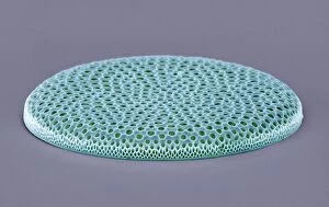
Background imageSingle Celled Collection: Coscinodiscus diatom, SEM

Coscinodiscus diatom, SEM
Coscinodiscus diatom. Coloured scanning electron micrograph (SEM) of a Coscinodiscus diatom (red). This is a marine diatom

Background imageSingle Celled Collection: Green alga, light micrograph

Green alga, light micrograph
Green alga. Differential interference contrast micrograph of the desmid green alga Euastrum didelta. Desmids are a common group of freshwater single-celled algae that have intricate cell walls. E

Background imageSingle Celled Collection: Fish killed by red tide

Fish killed by red tide. Dead fish killed as a result of a bloom of Karenia brevis. K breves is a microscopic single-celled marine dinoflagellate that blooms frequently along the Florida

Background imageSingle Celled Collection: Desmid alga, SEM

Desmid alga, SEM
Desmid. Coloured scanning electron micrograph (SEM) of a desmid. Desmids are a common type of green algae and are usually found in freshwater


All Professionally Made to Order for Quick Shipping

"Unveiling the Microscopic World: Captivating Single-Celled Organisms Under the SEM" Delicate and intricate, diatoms reveal their mesmerizing beauty under the scanning electron microscope (SEM). Their symmetrical frustules showcase nature's artistry at its finest. Witness the fascinating process of cell division in yeast cells as they multiply and propagate. The SEM captures this microscopic dance with remarkable detail. Behold the captivating artwork of Trypanosome protozoan, a single-celled organism that evokes both awe and curiosity. Its unique form is brought to life through artistic interpretation. Diatom alga, another marvel of nature, showcases its stunning structure when examined under the SEM lens. These tiny organisms play a crucial role in aquatic ecosystems worldwide. Protozoa, scavengers extraordinaire. Discover these single-celled wonders as they scavenge for particles and microorganisms or absorb nutrients from their environment – truly masters of survival. Acrosphaera radiolarian takes center stage with its intricate skeletal structure captured by SEM imaging techniques. Marvel at this ancient marine creature frozen in time. Journey back millions of years as fossilized diatoms come into focus under the SEM lens, offering glimpses into Earth's past and providing valuable insights for scientific research today. Diatom frustule - an architectural masterpiece on a microscopic scale. Explore these silica-based shells that protect diatoms while showcasing an array of breathtaking patterns through SEM imagery. Oxytricha ciliate protozoan reveals its astonishing complexity when observed using advanced microscopy techniques like SEM C019 / 0253 – unlocking secrets hidden within this enigmatic world. Exploring single-celled organisms through scanning electron microscopy opens up a whole new dimension where beauty meets science; revealing intricacies unseen by our naked eyes but essential to understanding life's diversity on our planet.