Fraud Blocker Skip to main content

Respiratory Collection (#4)

"The Marvels of the Respiratory System: Exploring the Gateway to Life" Diagram of the Lungs and Bronchial Tubes

Background imageRespiratory Collection: Physical fitness test C014 / 1107

Physical fitness test C014 / 1107
Physical fitness test. Athlete riding an exercise bike while his oxygen consumption is measured with a face mask. Electrodes on his chest monitor his heart rate

Background imageRespiratory Collection: Physical fitness test C014 / 1102

Physical fitness test C014 / 1102
Physical fitness test. Athlete riding an exercise bike while his oxygen consumption is measured with a face mask. Electrodes on his chest monitor his heart rate

Background imageRespiratory Collection: Tuberculosis X-ray diagnosis

Tuberculosis X-ray diagnosis
MODEL RELEASED. Tuberculosis (TB) X-ray diagnosis. Close-up of an X-ray of a patients lungs being examined by medical personnel for signs of TB

Background imageRespiratory Collection: Physical fitness test C014 / 1100

Physical fitness test C014 / 1100
Physical fitness test. Athlete undergoing a physical examination. Here his lung capacity is being tested using a spirometer, which measures the volume and speed of air that he can exhale and inhale

Background imageRespiratory Collection: Grape leaf stoma, SEM C014 / 4740

Grape leaf stoma, SEM C014 / 4740
Grape leaf stoma. Coloured scanning electron micrograph (SEM) of a closed stoma (centre-right) on a leaf from a grape (Vitis sp.) vine

Background imageRespiratory Collection: CPR first aid technique, artwork

CPR first aid technique, artwork
CPR first aid technique. Artwork showing the 2nd of 6 steps used in the first aid technique of cardiopulmonary resuscitation (CPR)

Background imageRespiratory Collection: Mitochondrial structure, artwork

Mitochondrial structure, artwork
Mitochondrial structure. Computer artwork of a section through a mitochondrion, showing the internal structure and a loop of mitochondrial DNA (deoxyribonucleic acid, mtDNA)

Background imageRespiratory Collection: Heart-lungs circulatory system, artwork

Heart-lungs circulatory system, artwork
Heart-lungs circulatory system, computer artwork. The heart (centre) is a hollow muscle that pumps blood around the body. The two branches of the pulmonary artery (red)

Background imageRespiratory Collection: Heart-lung circulatory system, artwork

Heart-lung circulatory system, artwork
Heart-lung circulatory system, computer artwork. The heart (centre) is a hollow muscle that pumps blood around the body. The two branches of the pulmonary artery (red)

Background imageRespiratory Collection: Lung blood vessel, artwork

Lung blood vessel, artwork
Lung blood vessels, computer artwork. Shown are the lung blood vessels in side view. The branches of the pulmonary artery (red) which carry deoxygenated blood from the heart to the lungs

Background imageRespiratory Collection: Carbon dioxide molecules C016 / 8495

Carbon dioxide molecules C016 / 8495
Carbon dioxide molecules. Computer artwork showing the structure of a molecule of carbon dioxide. Carbon dioxide is a colourless gas that occurs naturally in the atmosphere

Background imageRespiratory Collection: Carbon dioxide molecule C016 / 8494

Carbon dioxide molecule C016 / 8494
Carbon dioxide molecule. Computer artwork showing the structure of a molecule of carbon dioxide. Carbon dioxide is a colourless gas that occurs naturally in the atmosphere

Background imageRespiratory Collection: Carbon dioxide molecule C016 / 8493

Carbon dioxide molecule C016 / 8493
Carbon dioxide molecule. Computer artwork showing the structure of a molecule of carbon dioxide. Carbon dioxide is a colourless gas that occurs naturally in the atmosphere

Background imageRespiratory Collection: Influenza virus, artwork C016 / 8345

Influenza virus, artwork C016 / 8345
This image may not be used in educational posters Influenza virus. Cut-away computer artwork of an influenza (flu) virus particle (virion)

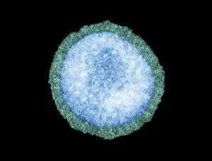
Background imageRespiratory Collection: Influenza virus particle, TEM

Influenza virus particle, TEM
Influenza virus particle, coloured transmission electron micrograph (TEM). The virus consists of ribonucleic acid (RNA), surrounded by a nucleocapsid (light green) and a lipid envelope (dark green)

Background imageRespiratory Collection: Heart-lungs circulatory system

Heart-lungs circulatory system, computer artwork. The heart (centre) is a hollow muscle that pumps blood around the body. The two branches of the pulmonary artery (red)

Background imageRespiratory Collection: RSV virus fusion glycoprotein C015 / 6689

RSV virus fusion glycoprotein C015 / 6689
RSV virus fusion glycoprotein, molecular model. This is a fusion glycoprotein of human respiratory syncytial virus (RSV). This glycoprotein is used by the virus to invade host cells

Background imageRespiratory Collection: RSV virus fusion glycoprotein C015 / 6688

RSV virus fusion glycoprotein C015 / 6688
RSV virus fusion glycoprotein, molecular model. This is a fusion glycoprotein of human respiratory syncytial virus (RSV). This glycoprotein is used by the virus to invade host cells

Background imageRespiratory Collection: Oxidoreductase enzyme complex C015 / 6554

Oxidoreductase enzyme complex C015 / 6554
Oxidoreductase enzyme complex, molecular model. This is the membrane-bound domain formed from of a complex of NADH-quinone oxidoreductase subunits

Background imageRespiratory Collection: Smog from wildfires, Tomsk C015 / 5889

Smog from wildfires, Tomsk C015 / 5889
Smog. View of the city of Tomsk, Russia, through a heavy smog caused by local wild fires. Photographed in July 2012

Background imageRespiratory Collection: Oxidoreductase enzyme complex C015 / 6553

Oxidoreductase enzyme complex C015 / 6553
Oxidoreductase enzyme complex, molecular model. This is the membrane-bound domain formed from of a complex of NADH-quinone oxidoreductase subunits

Background imageRespiratory Collection: Culex mosquito larva C017 / 8352

Culex mosquito larva C017 / 8352
Culex mosquito larva, with a breathing tube, on the waters surface. Mosquito larvae have a well-developed head (upper right) with mouth brushes used for feeding and propulsion

Background imageRespiratory Collection: Human anatomy, 1823 C017 / 8057

Human anatomy, 1823 C017 / 8057
Human anatomy. 19th-century illustrations showing the anatomy of a human skull (upper left), brain and spinal column (upper centre), chest and abdominal organs (lungs, heart, liver, stomach)

Background imageRespiratory Collection: Pneumothorax, X-ray C017 / 7966

Pneumothorax, X-ray C017 / 7966
Pneumothorax. X-ray of the chest of a 22 year old male patient with a pneumothorax, or collapsed lung. The left lung (right on X-ray) has collapsed due to a build-up of air (solid grey)

Background imageRespiratory Collection: Tuberculosis campaign, early 20th century C017 / 7885

Tuberculosis campaign, early 20th century C017 / 7885
Tuberculosis campaign, early 20th century. Doctor using a stethoscope to check a boys lungs for signs of the respiratory bacterial disease tuberculosis (TB)

Background imageRespiratory Collection: Lung disease, X-ray C017 / 7817

Lung disease, X-ray C017 / 7817
Lung disease. X-ray of the chest of a patient with chronic obstructive pulmonary disease (COPD). This is a collection of diseases that includes emphysema and chronic bronchitis

Background imageRespiratory Collection: Gapeworm, SEM C016 / 3085

Gapeworm, SEM C016 / 3085
Gapeworm. Coloured scanning electron micrograph of a female gapeworm (Syngamus trachea) from an owl. Gapeworms are parasitic nematode worms that infect the trachea (windpipe) of certain birds

Background imageRespiratory Collection: Gapeworm, SEM C016 / 3084

Gapeworm, SEM C016 / 3084
Gapeworm. Coloured scanning electron micrograph of a female gapeworm (Syngamus trachea) from an owl. Gapeworms are parasitic nematode worms that infect the trachea (windpipe) of certain birds

Background imageRespiratory Collection: Gapeworm head, SEM C016 / 3082

Gapeworm head, SEM C016 / 3082
Gapeworm head. Coloured scanning electron micrograph of the head of a female gapeworm (Syngamus trachea) from an owl. Gapeworms are parasitic nematode worms that infect the trachea (windpipe)

Background imageRespiratory Collection: Gapeworm head, SEM C016 / 3083

Gapeworm head, SEM C016 / 3083
Gapeworm head. Coloured scanning electron micrograph of the head of a female gapeworm (Syngamus trachea) from an owl. Gapeworms are parasitic nematode worms that infect the trachea (windpipe)

Background imageRespiratory Collection: NADH dehydrogenase molecule, artwork C014 / 0009

NADH dehydrogenase molecule, artwork C014 / 0009
NADH dehydrogenase molecule. Computer artwork showing the structure of a molecule of NADH dehydrogenase (NADH:ubiquinone reductase or Complex I)

Background imageRespiratory Collection: Obesity clinic C013 / 8716

Obesity clinic C013 / 8716
MODEL RELEASED. Obesity clinic. Patient using a walking aid. The patient is wearing a tube to provide her with oxygen-enriched air

Background imageRespiratory Collection: Obesity clinic C013 / 8715

Obesity clinic C013 / 8715
MODEL RELEASED. Obesity clinic. Physiotherapist talking with a patient. The patient is wearing a tube to provide her with oxygen-enriched air

Background imageRespiratory Collection: Bird flu virus particle, artwork C013 / 4650

Bird flu virus particle, artwork C013 / 4650
Bird flu virus particle. Computer artwork showing the internal and external structures of an influenza (flu) A subtype H5N1 (bird flu) virus particle (virion)

Background imageRespiratory Collection: Stomata of Lavendula Dentata, SEM

Stomata of Lavendula Dentata, SEM
Open and closed stomata on a lavender leaf (Lavendula dentata), coloured scanning electron micrograph (SEM). Stomata are pores that open and close in order to regulate gas exchange in a plant

Background imageRespiratory Collection: Mine rescue team wearing breathing apparatus

Mine rescue team wearing breathing apparatus
A mine rescue team wearing new breathing apparatus set off into the mine by truck. circa 1911

Background imageRespiratory Collection: Men testing breathing apparatus

Men testing breathing apparatus
A group of men testing some breathing apparatus, possibly for diving or for use down a mine. circa 1911

Background imageRespiratory Collection: Sea Cucumber (Holothuroidea sp. ) adult, extruding white sticky respiratory tubules in defensive

Sea Cucumber (Holothuroidea sp. ) adult, extruding white sticky respiratory tubules in defensive action at night
Sea Cucumber (Holothuroidea sp.) adult, extruding white sticky respiratory tubules in defensive action at night, Gam Island, Raja Ampat, West Papua, New Guinea, Indonesia

Background imageRespiratory Collection: Leopard Sea Cucumber (Bohadschia argus) adult, extruding white sticky respiratory tubules in

Leopard Sea Cucumber (Bohadschia argus) adult, extruding white sticky respiratory tubules in defensive action, Pulau Nailaka, Banda, near Ambon Island, Maluku Islands, Banda Sea, Indonesia

Background imageRespiratory Collection: Mantle of a giant clam

Mantle of a giant clam (Tridacna gigas). The clams mantle tissues contain symbiotic singled- celled dinoflagellate algae (zooxanthellae), which provide it with nutrition and its colour

Background imageRespiratory Collection: Silk moth caterpillar breathing pore

Silk moth caterpillar breathing pore
Silk moth caterpillar air pore. Polarised light micrograph of a spiracle (air pore, upper centre) and its attached tracheae (air passages) from a silk moth caterpillar (Bombyx mori)

Background imageRespiratory Collection: Breathing tube on a fruit flys pupa, SEM

Breathing tube on a fruit flys pupa, SEM
Breathing tube on a fruit flys pupa, coloured scanning electron micrograph (SEM). This structure is called a spiracle. This fruit fly is Drosophila melanogaster (wild type Oregon R)

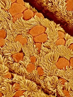
Background imageRespiratory Collection: Lung lining, SEM

Lung lining, SEM
Lung lining. Coloured scanning electron micrograph (SEM) of mucus-producing cells (orange, round) and cilia (yellow) lining a bronchus (lung airway)

Background imageRespiratory Collection: Lung, X-ray

Lung, X-ray
Lung. Coloured bronchography (X-ray) of a healthy human lung. A contrast medium has been added to show the network of airways (green) in the right lung

Background imageRespiratory Collection: Lung cells, light micrograph

Lung cells, light micrograph
Lung cells. Light micrograph of a section through the wall of a bronchus in the lungs. This is the upper layer of cells (the epithelium). The cell nuclei (round, dark) are across bottom

Background imageRespiratory Collection: Skeleton with stethoscope

Skeleton with stethoscope. The ear pieces of the stethoscope are wrapped around the skeletons neck, and the diaphragm is tucked into the ribcage

Background imageRespiratory Collection: Adenovirus particles and bacterium, TEM

Adenovirus particles and bacterium, TEM
Adenovirus particles and bacterium. Coloured transmission electron micrograph (TEM) of adenovirus particles (green) with a gut bacterium (orange)

Background imageRespiratory Collection: Respiratory syncytial virus, TEM

Respiratory syncytial virus, TEM
Respiratory syncytial virus (RSV) particles, coloured transmission electron micrograph (TEM). This pneumovirus, a type of paramyxovirus


All Professionally Made to Order for Quick Shipping

"The Marvels of the Respiratory System: Exploring the Gateway to Life" Diagram of the Lungs and Bronchial Tubes: Unveiling the intricate network within our bodies that facilitates every breath we take, this diagram showcases the lungs and bronchial tubes, highlighting their vital role in oxygenating our bloodstream. Diagram of the Heart, Lungs, and Windpipe: Witnessing a symphony of life-sustaining organs working harmoniously together, this diagram depicts the heart pumping blood while being surrounded by the lungs and windpipe – a testament to our body's remarkable respiratory system. Illustration of Human Respiratory System: Embark on an anatomical journey through our respiratory system as this illustration reveals its various components such as oral cavity, nasal cavity, larynx, trachea, bronchus, and lungs – each playing a crucial part in ensuring efficient respiration. Tension Pneumothorax X-ray: Peering into medical imaging technology's window into diagnosis and treatment strategies for collapsed lungs caused by tension pneumothorax - an urgent condition requiring immediate attention to restore normal breathing patterns. English Oak Leaf Pores SEM Image: Delving into nature's wonders at microscopic levels reveals mesmerizing images showcasing English oak leaf pores under scanning electron microscopy (SEM), reminding us how even plants have their unique respiratory systems. Cystic Fibrosis: Unmasking a Genetic Challenge Exploring cystic fibrosis - a genetic disorder affecting lung function - sheds light on ongoing research efforts aimed at improving treatments for individuals facing challenges with their respiratory health from an early age. Konstantin Buteyko: Soviet Doctor Revolutionizing Breathing Techniques Discovering how Dr. Konstantin Buteyko revolutionized breathing techniques offers insights into his innovative methods that have helped countless individuals manage asthma symptoms and enhance overall respiratory well-being. Paramyxovirus Particles TEM Image.