Fraud Blocker Skip to main content

Pathogenic Collection (#4)

Pathogenic microorganisms are microscopic entities that pose a significant threat to human health

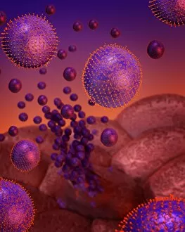
Background imagePathogenic Collection: Flu virus infection, conceptual image

Flu virus infection, conceptual image. Computer artwork showing influenza virus particles (round) in the respiratory tract

Background imagePathogenic Collection: Avian polyomavirus capsid

Avian polyomavirus capsid, molecular model. This virus, one of a range named for their potential to cause multiple tumours, infects birds. Discovered in budgerigars in 1981, it is often fatal

Background imagePathogenic Collection: Cytoplasmic polyhedrosis virus capsid

Cytoplasmic polyhedrosis virus capsid, molecular model. Part of the Cypovirus genus and invariably fatal, this insect virus is transmitted by contamination of leaves eaten (examples include silkworms)

Background imagePathogenic Collection: Theilers encephalomyelitis virus capsid

Theilers encephalomyelitis virus capsid, molecular model. This virus, which causes brain and spinal cord inflammation in mice, is used in research

Background imagePathogenic Collection: Coronavirus, artwork F007 / 0229

Coronavirus, artwork F007 / 0229
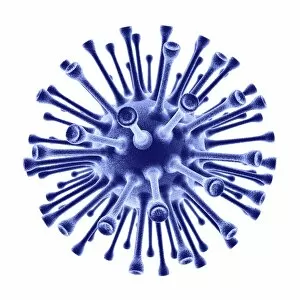
Human coronavirus. Computer artwork of a Human coronavirus particle. Coronaviruses primarily infect the upper respiratory and gastrointestinal tract and can cause the common cold

Background imagePathogenic Collection: Coronavirus, artwork F007 / 0222

Coronavirus, artwork F007 / 0222
Human coronavirus. Computer artwork of a Human coronavirus particle. Coronaviruses primarily infect the upper respiratory and gastrointestinal tract and can cause the common cold

Background imagePathogenic Collection: Coronavirus, artwork F007 / 0220

Coronavirus, artwork F007 / 0220
Human coronavirus. Computer artwork of a Human coronavirus particle. Coronaviruses primarily infect the upper respiratory and gastrointestinal tract and can cause the common cold

Background imagePathogenic Collection: Coronavirus, artwork F007 / 0211

Coronavirus, artwork F007 / 0211
Human coronavirus. Computer artwork of a Human coronavirus particle. Coronaviruses primarily infect the upper respiratory and gastrointestinal tract and can cause the common cold

Background imagePathogenic Collection: Tobacco necrosis virus capsid

Tobacco necrosis virus capsid, molecular model. This plant virus infects a wide rage of plants, including the tobacco plant for which it is named. The virus causes tissue death (necrosis)

Background imagePathogenic Collection: Genetic evolution of flu virus, artwork C017 / 0786

Genetic evolution of flu virus, artwork C017 / 0786
Genetic evolution of influenza (flu) virus. Artwork showing how reassortment of the influenza virus genome led to the evolution of the H7N9 virus in China

Background imagePathogenic Collection: Grapevine fanleaf virus capsid

Grapevine fanleaf virus capsid, molecular model. This plant virus is named for its infection of grape vines. It is transmitted by the nematode worm Xiphinema index

Background imagePathogenic Collection: SV40 virus capsid, molecular model F006 / 9508

SV40 virus capsid, molecular model F006 / 9508
SV40 virus capsid, molecular model. Simian virus 40 (SV40) is found in monkeys such as Rhesus monkeys and macaques. Potentially tumour-causing, it is used in laboratory research and in vaccines

Background imagePathogenic Collection: Human poliovirus, molecular model F006 / 9434

Human poliovirus, molecular model F006 / 9434
Human poliovirus particle. Molecular model of the capsid of the human poliovirus. The capsid is a protein coat that encloses the viruss genetic information (genome), stored as RNA (ribonucleic acid)

Background imagePathogenic Collection: VEE equine encephalitis virus capsid

VEE equine encephalitis virus capsid
Venezuelan equine encephalitis virus capsid, molecular model. This mosquito-borne virus can kill horses and other equine species, causing brain and spinal cord inflammation

Background imagePathogenic Collection: Poliovirus particle F006 / 9306

Poliovirus particle F006 / 9306
Poliovirus particle, molecular model

Background imagePathogenic Collection: Semliki forest virus capsid F006 / 9297

Semliki forest virus capsid F006 / 9297
Semliki forest virus capsid, molecular model. This virus, named for the forest in Uganda where it was identified, is spread by the bite of mosquitoes. It can infect both humans and animals

Background imagePathogenic Collection: Human poliovirus, molecular model F006 / 9289

Human poliovirus, molecular model F006 / 9289
Human poliovirus particle. Molecular model of the capsid of the human poliovirus. The capsid is a protein coat that encloses the viruss genetic information (genome), stored as RNA (ribonucleic acid)

Background imagePathogenic Collection: H1N1 flu virus particle, artwork F006 / 9186

H1N1 flu virus particle, artwork F006 / 9186
H1N1 flu virus particle. Computer artwork of an H1N1 influenza A (flu) virus particle (virion). In the particles lipid envelope (purple) are two types of protein spike

Background imagePathogenic Collection: Murine minute virus capsid

Murine minute virus capsid, molecular model. This parvovirus infects mice, its only known natural host. It is highly infectious, transmitted through the nose and mouth

Background imagePathogenic Collection: SV40 virus capsid, molecular model C018 / 7904

SV40 virus capsid, molecular model C018 / 7904
SV40 virus capsid, molecular model. Simian virus 40 (SV40) is found in monkeys such as Rhesus monkeys and macaques. Potentially tumour-causing, it is used in laboratory research and in vaccines

Background imagePathogenic Collection: SV40 virus capsid, molecular model C018 / 7903

SV40 virus capsid, molecular model C018 / 7903
SV40 virus capsid, molecular model. Simian virus 40 (SV40) is found in monkeys such as Rhesus monkeys and macaques. Potentially tumour-causing, it is used in laboratory research and in vaccines

Background imagePathogenic Collection: Reovirus particles, TEM C016 / 9443

Reovirus particles, TEM C016 / 9443
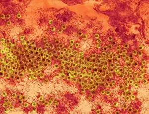
Reovirus particles. Coloured transmission electron micrograph (TEM) of numerous reovirus particles (virions, round). Reoviruses affect the gastrointestinal system and respiratory tract

Background imagePathogenic Collection: Pseudomonas aeruginosa bacterium, SEM C016 / 9442

Pseudomonas aeruginosa bacterium, SEM C016 / 9442
Pseudomonas aeruginosa bacterium, coloured transmission electron micrograph (TEM). P. aeruginosa is a Gram-negative rod-shaped bacterium found in soil and water

Background imagePathogenic Collection: White blood cell antigen presentation C016 / 9058

White blood cell antigen presentation C016 / 9058
White blood cell antigen presentation. Coloured scanning electron micrograph (SEM) showing the interaction between a macrophage (yellow) and a T helper lymphocyte (Th cell, green)

Background imagePathogenic Collection: Salmonella bacteria, artwork C016 / 7538

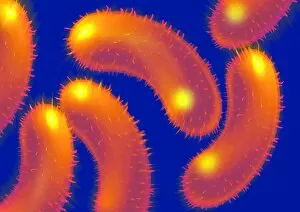
Salmonella bacteria, artwork C016 / 7538
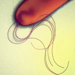
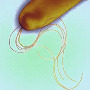
Salmonella bacteria, computer artwork. Salmonella sp. bacteria are gram-negative rod-shaped bacteria that have flagella (hair-like structures) that they use for locomotion

Background imagePathogenic Collection: Yellow fever virus particles, TEM C016 / 9468

Yellow fever virus particles, TEM C016 / 9468
Yellow fever virus particles. Transmission electron micrograph (TEM) of yellow fever virus particles (virions, yellow). Yellow fever is endemic in tropical Africa and South America

Background imagePathogenic Collection: Hepatitis B virus particle, artwork C016 / 9129

Hepatitis B virus particle, artwork C016 / 9129
Hepatitis B virus (HBV) particle, computer artwork. Each particle consists of an outer lipid envelope (green) that surrounds a protein nucleopcapsid (purple)

Background imagePathogenic Collection: Mycobacterium fortuitum bacteria C016 / 9424

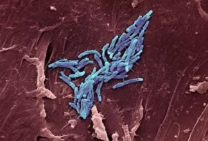
Mycobacterium fortuitum bacteria C016 / 9424
Mycobacterium fortuitum bacteria, coloured scanning electron micrograph (SEM). These bacteria (blue) are a non-tuberculosis strain of Mycobacterium

Background imagePathogenic Collection: Lassa virus particles, TEM C016 / 9409

Lassa virus particles, TEM C016 / 9409
Lassa virus particles. Transmission electron micrograph (TEM) of Lassa virus particles (virions, blue) amongst cell debris. This Arenavirus is the cause of Lassa fever

Background imagePathogenic Collection: Orientia tsutsugamushi bacterium C016 / 9431

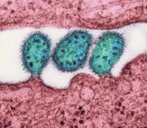
Orientia tsutsugamushi bacterium C016 / 9431
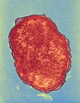
Orientia tsutsugamushi bacterium. Transmission electron micrograph (TEM) of a section through an Orientia tsutsugamushi bacterium. O

Background imagePathogenic Collection: HIV particles, artwork C016 / 9141

HIV particles, artwork C016 / 9141
HIV particles, computer artwork. HIV (human immunodeficiency virus) causes AIDS (acquired immune deficiency syndrome). The virus consists of an RNA (ribonucleic acid) genome (pink)

Background imagePathogenic Collection: Hepatitis B virus particle, artwork C016 / 9126

Hepatitis B virus particle, artwork C016 / 9126
Hepatitis B virus (HBV) particle, computer artwork. Each particle consists of an outer lipid envelope (green) that surrounds a protein nucleopcapsid (purple)

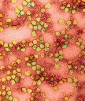
Background imagePathogenic Collection: Yellow fever virus particles, TEM C016 / 9469

Yellow fever virus particles, TEM C016 / 9469
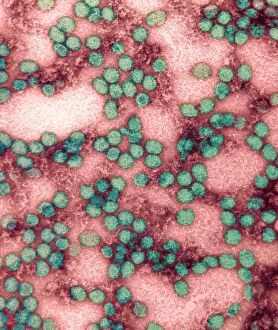
Yellow fever virus particles. Transmission electron micrograph (TEM) of yellow fever virus particles (virions, green). Yellow fever is endemic in tropical Africa and South America

Background imagePathogenic Collection: Pseudomonas aeruginosa bacterium, SEM C016 / 9441

Pseudomonas aeruginosa bacterium, SEM C016 / 9441
Pseudomonas aeruginosa bacterium, coloured transmission electron micrograph (TEM). P. aeruginosa is a Gram-negative rod-shaped bacterium found in soil and water

Background imagePathogenic Collection: Human adenovirus 36, artwork C016 / 8966

Human adenovirus 36, artwork C016 / 8966
Human adenovirus 36. Computer artwork of human adenovirus 36 (HAdV-36), showing the surface structure of the viruss outer protein coat (capsid)

Background imagePathogenic Collection: SARS virus particles, TEM C016 / 9445

SARS virus particles, TEM C016 / 9445
SARS virus particles. Coloured transmission electron micrograph (TEM) of a section through a tissue sample infected with numerous SARS coronavirus particles (virions, dark, round)

Background imagePathogenic Collection: Polio virus particles, TEM C016 / 9436

Polio virus particles, TEM C016 / 9436
Poliovirus particles. Coloured transmission electron micrograph (TEM) of poliovirus particles (round). Poliovirus infects children causing the disease poliomyelitis, or infantile paralysis

Background imagePathogenic Collection: Acute haemorrhagic conjunctivitis, TEM C016 / 9387

Acute haemorrhagic conjunctivitis, TEM C016 / 9387
Acute haemorrhagic conjunctivitis. Transmission electron micrograph (TEM) of a sample of tissue from the conjunctiva of a human eye that is infected by enterovirus particles (purple)

Background imagePathogenic Collection: Eastern equine encephalitis, TEM C016 / 9390

Eastern equine encephalitis, TEM C016 / 9390
Eastern equine encephalitis. Transmission electron micrograph (TEM) of a section through tissue from a human central nervous system (CNS)

Background imagePathogenic Collection: MRSA bacteria, SEM C016 / 9422

MRSA bacteria, SEM C016 / 9422
MRSA bacteria. Coloured scanning electron micrograph (SEM) of methicillin-resistant Staphylococcus aureus (MRSA, round) bacteria

Background imagePathogenic Collection: Colorado tick fever virus infection, TEM C016 / 9384

Colorado tick fever virus infection, TEM C016 / 9384
Colorado tick fever virus infection. Transmission electron micrograph (TEM) of human tissue infected by Colorado tick fever virus (CTFV) particles (virions, dark circles)

Background imagePathogenic Collection: Syphilis bacterium, TEM C016 / 9461

Syphilis bacterium, TEM C016 / 9461
Syphilis bacteria. Transmission electron micrograph (TEM) of a Treponema pallidum spirochaete bacterium. T. pallidum is the bacteria that is responsible for the sexually transmitted infection syphilis

Background imagePathogenic Collection: Helicobacter pylori bacteria, SEM C016 / 9135

Helicobacter pylori bacteria, SEM C016 / 9135
Helicobacter pylori bacteria. Coloured scanning electron micrograph (SEM) of Helicobacter pylori bacteria (yellow) in the stomach. H

Background imagePathogenic Collection: Helicobacter pylori bacteria, SEM C016 / 9134

Helicobacter pylori bacteria, SEM C016 / 9134
Helicobacter pylori bacteria. Coloured scanning electron micrograph (SEM) of Helicobacter pylori bacteria (orange) in the stomach. H

Background imagePathogenic Collection: Swine flu virus particles, TEM C016 / 9399

Swine flu virus particles, TEM C016 / 9399
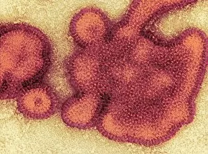
Swine flu virus particles. Transmission electron micrograph (TEM) of a section through H3N2 influenza A virus particles (virions)

Background imagePathogenic Collection: Swine flu virus particles, TEM C016 / 9406

Swine flu virus particles, TEM C016 / 9406
Influenza virus particles. Transmission electron micrograph (TEM) of influenza (flu) virus particles (virions). Each virion consists of ribonucleic acid (RNA, dark patches)

Background imagePathogenic Collection: Syphilis bacterium, TEM C016 / 9462

Syphilis bacterium, TEM C016 / 9462
Syphilis bacteria. Transmission electron micrograph (TEM) of a Treponema pallidum spirochaete bacterium. T. pallidum is the bacteria that is responsible for the sexually transmitted infection known

Background imagePathogenic Collection: St. Louis encephalitis virus particles C016 / 9453

St. Louis encephalitis virus particles C016 / 9453
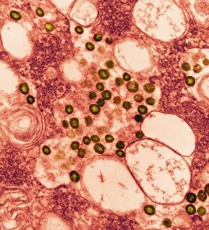
St. Louis encephalitis virus particles. Coloured transmission electron micrograph (TEM) of a section through a mosquito salivary gland containing numerous Saint Louis encephalitis virus particles


All Professionally Made to Order for Quick Shipping

Pathogenic microorganisms are microscopic entities that pose a significant threat to human health. From budding yeast cells to avian flu viruses, these tiny organisms have the potential to cause widespread diseases and infections. In the world of pathogens, the avian flu virus stands out as one of the most notorious culprits. Its ability to jump from birds to humans has sparked global concerns about pandemics and led scientists on a quest for effective prevention strategies. Neutrophils, our body's first line of defense against infection, play a crucial role in engulfing harmful bacteria like MRSA (methicillin-resistant Staphylococcus aureus). This SEM image captures this incredible process, showcasing how neutrophils work tirelessly to protect us from pathogenic invaders. Salmonella bacteria are another well-known pathogen responsible for causing foodborne illnesses worldwide. This SEM image provides an up-close look at their unique structure and highlights their capacity for survival in various environments. The HIV particle is infamous for its devastating impact on the immune system. Understanding its intricate structure has been instrumental in developing antiretroviral therapies that help manage this deadly virus. Anthrax cultures depicted in historical diagrams remind us of past outbreaks and highlight the importance of ongoing research into preventing bioterrorism threats associated with this highly infectious disease. Norovirus particles captured through TEM imaging reveal their distinctive shape and shed light on why they are so contagious. Responsible for many cases of gastroenteritis, noroviruses can spread rapidly within communities if proper hygiene measures aren't followed diligently. Tuberculosis bacteria continue to be a major global health concern due to their ability to infect millions each year. Efforts towards improved diagnostics and treatment options remain critical in combating this persistent pathogen. E. coli bacteria serve as both harmless inhabitants of our gut flora but also dangerous pathogens when certain strains acquire virulence factors. This diverse bacterium reminds us that not all E. coli strains are created equal.