Fraud Blocker Skip to main content

Nerves Collection (#9)

"Nerves: Unraveling the Intricacies of our Body's Communication System" Our bodies are a complex network of interconnected systems

Background imageNerves Collection: Artwork of vomiting mechanism in human body

Artwork of vomiting mechanism in human body
Vomiting. Illustration of the mechanism that causes vomiting in the human body. An impulse from the front of the brain has stimulated the vomiting centre (green) in the brainstem

Background imageNerves Collection: Vomiting, artwork

Vomiting, artwork
Vomiting. Computer artwork of synapses, or junctions, (cones) between nerve cells and the exterior wall of the stomach. During the vomit reflex, nerve impulses are sent from the brain to the stomach

Background imageNerves Collection: Somatic peripheral nervous system

Somatic peripheral nervous system
Peripheral nervous system. Computer artwork of the main nerves of the somatic part of the peripheral nervous system. The peripheral nervous system does not include the brain and spinal cord

Background imageNerves Collection: Coloured artwork of spinal column & spinal nerves

Coloured artwork of spinal column & spinal nerves
Spinal nerves. Computer-enhanced historical illustration of the human spinal cord and spinal nerves. Red and blue blood vessels & red muscles of the back & the upper arms can be seen

Background imageNerves Collection: Vomiting reflex

Vomiting reflex. Computer artwork of the vomiting reflex. The reflex starts when a message is sent to the vomiting centre of the brainstem (at base of brain, blue)

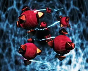
Background imageNerves Collection: Nanorobots in the brain

Nanorobots in the brain, conceptual image. In the future, it may be possible to introduce nanorobots into the human brain, where they will interact with biological neurones

Background imageNerves Collection: Prion protein plaque, computer artwork

Prion protein plaque, computer artwork
Prion protein plaque (right) surrounded by nerve cells (seen mainly on the left), computer artwork. Prions are infectious agents that cause a group of fatal neurodegenerative diseases

Background imageNerves Collection: Action of a beta blocker drug, artwork

Action of a beta blocker drug, artwork
Action of a beta blocker drug on nerve synapses, artwork. Beta blockers are used to treat cardiac arrhythmia (abnormal heartbeats) and hypertension (high blood pressure)

Background imageNerves Collection: Action of serotonin reuptake inhibitors

Action of serotonin reuptake inhibitors
Action of selective serotonin reuptake inhibitors (SSRIs) at a chemical synapse, computer artwork. Chemical synapses are the junctions between any two nerves (at right, blue) of the nervous system

Background imageNerves Collection: Neonatal reflex test

Neonatal reflex test
MODEL RELEASED. Neonatal reflex test. Doctor testing a newborn baby boys grasping reflex. This occurs when a finger or other object is placed in the babys palm

Background imageNerves Collection: Neural chip

Neural chip. Conceptual computer artwork of a neural network (centre) on a microchip. This could represent hardware which mimics such networks, or contains actual nerve cells

Background imageNerves Collection: Neuronal network

Neuronal network. Conceptual computer artwork of a neural network. This could represent the interior of a human brain or a biological computer memory which mimics such networks

Background imageNerves Collection: Schizophrenia, computer artwork

Schizophrenia, computer artwork
Schizophrenia. Computer artwork of areas of the brain thought to be involved in the disease schizophrenia. At centre are the basal ganglia (yellow)

Background imageNerves Collection: Shingles

Shingles. Artwork of varicella-zoster virus particles (virions, blue) during shingles. Each viron comprises an icosahedral (20-sided) capsid containing viral DNA

Background imageNerves Collection: Large intestine

Large intestine
Colon. Artwork showing the human colon (large intestine). The yellow fibres represent nerves, which control the involuntary muscular movements (peristalsis) of the colon

Background imageNerves Collection: Vision and the brain, MRI scan

Vision and the brain, MRI scan
Vision and the brain. Coloured Magnetic Resonance Imaging (MRI) scan of an axial section through the head of a 55 year old man, showing the eyes, optic nerves and brain

Background imageNerves Collection: Nerve cell synapses, computer artwork

Nerve cell synapses, computer artwork
Nerve cell synapses. Computer artwork of the chemical synapses between two nerve cells, or neurons, (one red and one blue)

Background imageNerves Collection: Synapse, computer artwork

Synapse, computer artwork
Synapses. Computer artwork of synapses, the junctions between the ends (blue, swollen) of two nerve cells (neurons). Nerve cells are responsible for passing information around the central nervous

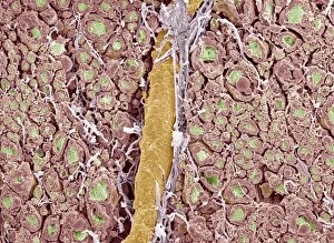
Background imageNerves Collection: Myelinated nerve fibres, SEM

Myelinated nerve fibres, SEM
Myelinated nerve fibres. Coloured scanning electron micrograph (SEM) of a section through a bundle of nerve fibres containing a blood vessel (orange)

Background imageNerves Collection: Neurovascular bundle, TEM

Neurovascular bundle, TEM
Neurovascular bundle. Coloured transmission electron micrograph (TEM) of a cross section through a neurovascular bundle (NVB)

Background imageNerves Collection: Nerve fibres, TEM

Nerve fibres, TEM
Nerve fibres. Coloured transmission electron micrograph (TEM) of a section through non- myelinated nerve fibres (blue). The fibres are arranged into bundles surrounded by the cytoplasm of Schwann

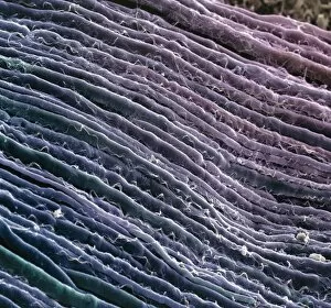
Background imageNerves Collection: Nerve fibres

Nerve fibres. Coloured scanning electron micrograph (SEM) of parallel myelinated nerve fibres in the spinal cord. Each fibre consists of a nerve cell axon, the output process of a nerve cell

Background imageNerves Collection: Coloured artwork of the human brain (side view)

Coloured artwork of the human brain (side view)
Brain. Computer-enhanced historical illustration of the sectioned human brain with cranial nerves, seen in side view. At top are surface folds of the cerebrum

Background imageNerves Collection: Blood vessels, light micrograph

Blood vessels, light micrograph
Blood vessels. Light micrograph of a section through tissue showing an artery (bottom left) and a vein (top right). Two nerve fibres (dark blue) are also seen

Background imageNerves Collection: Illustration of human muscle and movement systems

Illustration of human muscle and movement systems
Muscle and movement. Illustration of human muscle anatomy and movement systems. Movement depends on the interaction of multiple systems in the body, including muscle, bones and joints

Background imageNerves Collection: Kidneys, nerves and blood vessels

Kidneys, nerves and blood vessels
Kidneys, historical anatomical artwork. This dorsal (back) view shows the kidneys and associated nerves lying outside the peritoneum (membrane lining the abdominal cavity)

Background imageNerves Collection: Sympathetic nerves, lower body

Sympathetic nerves, lower body
Sympathetic nerves in the lower body. Historical anatomical artwork of a side view of a dissected human abdomen and chest, showing the origin and route of parts of the sympathetic nervous system

Background imageNerves Collection: Sympathetic nerves, upper body

Sympathetic nerves, upper body
Sympathetic nerves in the upper body. Historical anatomical artwork of a side view of a dissected human face, neck and chest, showing the origin and route of parts of the sympathetic nervous system

Background imageNerves Collection: Foot nerves

Foot nerves. Historical anatomical artwork of the nerves (white) of the sole of a human foot. Also shown are muscles (red), and ligaments, tendons and other connective tissues (grey)

Background imageNerves Collection: Leg nerves

Leg nerves. Historical anatomical artwork of the nerves (white) of the rear of a human lower leg. At left, the superficial (surface) nerves are seen and at right the deep nerves and muscles (red)

Background imageNerves Collection: Pelvic spinal nerves

Pelvic spinal nerves. Historical anatomical artwork of the spinal nerves (white) in the human pelvic region. This side view (front of the body at right)

Background imageNerves Collection: Abdominal spinal nerves

Abdominal spinal nerves. Historical anatomical artwork of the spinal nerves (white) in a human abdomen. This view, from the front

Background imageNerves Collection: Torso nerves

Torso nerves. Historical anatomical artwork of the nerves (white) of parts of a human torso. At lower frame, a side view of the abdomen (front of body at left) shows muscles (red)

Background imageNerves Collection: Arm nerves

Arm nerves. Historical anatomical artwork of the nerves (white) of the front (palm side) of a human forearm. At left, the superficial (surface) nerves are shown

Background imageNerves Collection: Twelfth cranial nerve

Twelfth cranial nerve (hypoglossal nerve, cranial nerve XII). Historical anatomical artwork of a side view of a dissected human neck showing veins (blue), arteries (red), muscles (red)

Background imageNerves Collection: Base of the brain

Base of the brain, historical anatomical artwork. The front of the brain is at top. The origin of the 12 cranial nerves is shown

Background imageNerves Collection: Brain

Brain, historical artwork. The top drawing shows the brain cut in-between its hemispheres. The front of the brain is on the left

Background imageNerves Collection: Knee reflex test

Knee reflex test
MODEL RELEASED. Knee reflex test. Physiotherapist using a testing mallet to perform a knee reflex test on a young child. The mallet is used to gently tap a specific area just below the childs knee

Background imageNerves Collection: Pain, conceptual computer artwork

Pain, conceptual computer artwork
Pain. Conceptual computer artwork of pain, where the site of pain is represented by a ball of barbed wire. Connected to this are the free nerve endings, which respond to mechanical

Background imageNerves Collection: Parkinsons disease

Parkinsons disease

Background imageNerves Collection: Cell death

Cell death. Computer-enhanced confocal light micrograph of cells in the retina of the eye undergoing programmed cell death (apoptosis)

Background imageNerves Collection: Female nervous system, artwork

Female nervous system, artwork
Female nervous system, computer artwork


All Professionally Made to Order for Quick Shipping

"Nerves: Unraveling the Intricacies of our Body's Communication System" Our bodies are a complex network of interconnected systems, and at the heart of it all lies our nervous system. From chakras to brain fibers, this captivating web controls every sensation we experience. Delving deeper into the mysteries, DTI MRI scans like C017/7099 and C017/7035 reveal intricate brain pathways that transmit signals throughout our body. These scans provide us with a glimpse into the fascinating world within. A diagram showcasing the human brain and spinal column reminds us of the delicate balance maintained by these vital structures, and is through these pathways that information flows seamlessly, allowing us to perceive and react to our surroundings. Even in lighthearted moments, such as a dentist cartoon by H. M. Bateman depicting cause and effect, we can see how nerves play their part in triggering reactions – both physical and emotional. The inner ear is another marvel worth exploring; its auditory canal, eardrum, semicircular canals, cochlea nerve, and eustachian tube work together harmoniously to process sound waves. Throughout history, scientists have tirelessly studied the nervous system's intricacies - an 18th-century depiction serves as a testament to humanity's enduring curiosity about this remarkable mechanism. Yet amidst all its wonders lie certain afflictions - migraines remind us that even they are sometimes falter under strain. Understanding facial nerves becomes crucial when dealing with conditions affecting expressions or sensations on our face. So next time you find yourself asking "Are you nervous?", take a moment to appreciate just how incredible your own nerve system truly is – for it enables you to feel emotions deeply while simultaneously coordinating countless bodily functions without conscious effort. Intricate yet resilient; sensitive yet powerful – our they can an awe-inspiring reminder of nature's brilliance woven within each one of us.