Fraud Blocker Skip to main content

Microbiology Collection

Microbiology, the fascinating world of tiny organisms and their impact on our lives

Background imageMicrobiology Collection: Tardigrade or Water Bear

Tardigrade or Water Bear
Water bear or tardigrade. Scanning electron micrograph (SEM) of a water bear (Echiniscus sp.) walking over spagnum moss. Water bears live in damp habitats such as moss or lichen

Background imageMicrobiology Collection: HeLa cells, light micrograph C017 / 8299

HeLa cells, light micrograph C017 / 8299
HeLa cells, multiphoton fluorescence micrograph (MFM). The cell nuclei, which contain the cells genetic information, are purple. Microtubules are blue and actin microfilaments are red

Background imageMicrobiology Collection: stem cell

stem cell
microscopic image

Background imageMicrobiology Collection: Picture No. 11675590

Picture No. 11675590
Macro Photograph. Date:

Background imageMicrobiology Collection: Neutrophil engulfing MRSA, SEM C018 / 8596

Neutrophil engulfing MRSA, SEM C018 / 8596
Neutrophil engulfing MRSA. Coloured scanning electron micrograph (SEM) of a neutrophil white blood cell (green) engulfing methicillin-resistant Staphylococcus aureus bacteria (MRSA, pink)

Background imageMicrobiology Collection: HeLa cells, light micrograph C017 / 8298

HeLa cells, light micrograph C017 / 8298
HeLa cells, multiphoton fluorescence micrograph (MFM). The cell nuclei, which contain the cells genetic information, are blue. Golgi bodies, which modify and package proteins, are orange

Background imageMicrobiology Collection: Yoghurt bacteria

Yoghurt bacteria. Coloured scanning electron micrograph (SEM) of the bacteria Streptococcus thermophilus (spherical) and Lactobacillus bulgar- icus (rod-shaped) in live yoghurt. S

Background imageMicrobiology Collection: Water bear, light micrograph C016 / 8581

Water bear, light micrograph C016 / 8581
Water bear, differential interference contrast (DIC) light micrograph. Water bears, or tardigrades, (phylum Tardigrada) are small, water-dwelling

Background imageMicrobiology Collection: Louis Pasteur. Artist: Edelfelt, Albert Gustaf Aristides (1854-1905)

Louis Pasteur. Artist: Edelfelt, Albert Gustaf Aristides (1854-1905)
Louis Pasteur. Found in the collection of Musee d Orsay, Paris

Background imageMicrobiology Collection: Microscopic view of human respiratory syncytial virus

Microscopic view of human respiratory syncytial virus (RSV). RSV causes respiratory tract infection of the lungs and breathing passages

Background imageMicrobiology Collection: Infections spread by sneezing, artwork C013 / 5949

Infections spread by sneezing, artwork C013 / 5949
Infections spread by sneezing. Computer artwork showing virus particles (virions, blue) and bacteria (rod-shaped) in the spray produced by someone when they sneeze

Background imageMicrobiology Collection: Cup lichen (Cladonia floerkeana)

Cup lichen (Cladonia floerkeana)
Lichen fruiting bodies (red). This lichen species is Cladonia floerkeana, found on heaths and moorlands. Lichens are formed from a symbiotic association between a fungus and an alga

Background imageMicrobiology Collection: The Research. Artist: Sorolla y Bastida, Joaquin (1863-1923)

The Research. Artist: Sorolla y Bastida, Joaquin (1863-1923)
The Research. Found in the collection of Museo Sorolla, Madrid

Background imageMicrobiology Collection: Illustration of response to infection after vaccination, involving microbe, antigens, antibody, plas

Illustration of response to infection after vaccination, involving microbe, antigens, antibody, plas

Background imageMicrobiology Collection: Embryo development 24-36 hours after fertilization

Embryo development 24-36 hours after fertilization

Background imageMicrobiology Collection: Microscopic view of chlamydia

Microscopic view of chlamydia. Chlamydia is a common sexually transmitted disease (STD) caused by a bacterium

Background imageMicrobiology Collection: Flu virus particle, artwork F008 / 3245

Flu virus particle, artwork F008 / 3245
Flu virus particle, computer artwork. In the virus envelope are two types of protein spike, haemagglutinin (H, pink) and neuraminidase (N, orange), which determine the strain of virus

Background imageMicrobiology Collection: Plankton, artwork C016 / 3447

Plankton, artwork C016 / 3447
Plankton. Artwork of plankton as viewed under a magnifying glass

Background imageMicrobiology Collection: Salmonella bacteria, artwork C013 / 8818

Salmonella bacteria, artwork C013 / 8818
Salmonella bacteria, computer artwork. Salmonella sp. bacteria are gram-negative rod-shaped bacteria that have flagella (hair-like structures) that they use for locomotion

Background imageMicrobiology Collection: Foraminifera, SEM

Foraminifera, SEM
Foraminifera. Coloured scanning electron micrograph (SEM) of a selection of foraminiferans, including Oolina hexagona (bottom right) and a number of Lagena sp

Background imageMicrobiology Collection: Water bear, SEM C016 / 9084

Water bear, SEM C016 / 9084
Water bear (Echiniscus granulatus). Coloured scanning electron micrograph (SEM) showing the underside of a water bear, or tardigrade (phylum Tardigrada)

Background imageMicrobiology Collection: Oxytricha ciliate protozoan, SEM C019 / 0253

Oxytricha ciliate protozoan, SEM C019 / 0253
Oxytricha sp. ciliate protozoan, coloured scanning electron micrograph (SEM). Oxytricha is a tiny single-celled aquatic organism

Background imageMicrobiology Collection: Polyoma BK virus, artwork C013 / 7465

Polyoma BK virus, artwork C013 / 7465
Computer artwork of the capsid of a polyoma BK virus. This polyomavirus is common in the urinary tract of adults, where it lives without harming its host

Background imageMicrobiology Collection: Microscope by Adams of London

Microscope by Adams of London
5311766 Microscope by Adams of London; (add.info.: Illustration of a microscope by Adams of London. Dated circa 1788.); Universal History Archive/UIG

Background imageMicrobiology Collection: Robert Koch working in his laboratory, 1850

Robert Koch working in his laboratory, 1850
3359517 Robert Koch working in his laboratory, 1850; (add.info.: Robert Koch (1843-1910) a German physician working in his laboratory. Dated 19th Century.); Universal History Archive/UIG

Background imageMicrobiology Collection: Bacteriological laboratory, France, 1890s. Chromolithograph c1900

Bacteriological laboratory, France, 1890s. Chromolithograph c1900
527488 Bacteriological laboratory, France, 1890s. Chromolithograph c1900.; Universal History Archive/UIG

Background imageMicrobiology Collection: Head of the Anopheles Mosquito showing the female mouthparts. Engraving, 1912

Head of the Anopheles Mosquito showing the female mouthparts. Engraving, 1912
527390 Head of the Anopheles Mosquito showing the female mouthparts. Engraving, 1912.; (add.info.: The bite of the female of this species can transmit the Malaria parasite)

Background imageMicrobiology Collection: An Louis Pasteur experiment, 19th century (engraving)

An Louis Pasteur experiment, 19th century (engraving)
3733827 An Louis Pasteur experiment, 19th century (engraving); (add.info.: Engraving depicting an Louis Pasteur experiment demonstrating that fermentation)

Background imageMicrobiology Collection: Commemorative medal depicting Louis Pasteur

Commemorative medal depicting Louis Pasteur
3733759 Commemorative medal depicting Louis Pasteur; (add.info.: Commemorative medal depicting Louis Pasteur (1822-1895) a French biologist

Background imageMicrobiology Collection: Three pillar microscope

Three pillar microscope
5311760 Three pillar microscope; (add.info.: Illustration of a Three pillar microscope of the type produced by Edmund Culpeper (1660-1738) circa 1730.); Universal History Archive/UIG

Background imageMicrobiology Collection: Louis Pasteur, 19th century (engraving)

Louis Pasteur, 19th century (engraving)
5310031 Louis Pasteur, 19th century (engraving); (add.info.: Portrait of Louis Pasteur (1822-1895) a French biologist, microbiologist and chemist. Dated 19th century.); Universal History Archive/UIG

Background imageMicrobiology Collection: Compound microscope

Compound microscope
5311762 Compound microscope; (add.info.: Illustration depicting a compound microscope by Adams of London. Dated circa 1788.); Universal History Archive/UIG

Background imageMicrobiology Collection: Louis Pasteur's first apparatus for cooling and fermenting wort

Louis Pasteur's first apparatus for cooling and fermenting wort
3733830 Louis Pasteur's first apparatus for cooling and fermenting wort.; (add.info.: Engraving depicting Louis Pasteur's first apparatus for cooling)

Background imageMicrobiology Collection: Louis Pasteur, 19th century (engraving)

Louis Pasteur, 19th century (engraving)
3733755 Louis Pasteur, 19th century (engraving); (add.info.: Portrait of Louis Pasteur (1822-1895) a French biologist, microbiologist

Background imageMicrobiology Collection: Louis Pasteur's method of observing sealed culture samples

Louis Pasteur's method of observing sealed culture samples
5310078 Louis Pasteur's method of observing sealed culture samples; (add.info.: Engraving depicting Louis Pasteur's method of observing sealed culture samples)

Background imageMicrobiology Collection: An Louis Pasteur experiment

An Louis Pasteur experiment
3733828 An Louis Pasteur experiment.; (add.info.: Engraving depicting an Louis Pasteur experiment demonstrating that fermentation and putrefaction are caused by air-borne organisms)

Background imageMicrobiology Collection: The exterior of the Ecole Normale Superieure, 19th century (engraving)

The exterior of the Ecole Normale Superieure, 19th century (engraving)
3733826 The exterior of the Ecole Normale Superieure, 19th century (engraving); (add.info.: Illustration depicting the exterior of the Ecole Normale Superieure, Paris)

Background imageMicrobiology Collection: Louis Pasteur's first apparatus for cooling and fermenting wort

Louis Pasteur's first apparatus for cooling and fermenting wort
3733829 Louis Pasteur's first apparatus for cooling and fermenting wort.; (add.info.: Engraving depicting Louis Pasteur's first apparatus for cooling)

Background imageMicrobiology Collection: Louis Pasteur, 19th century (engraving)

Louis Pasteur, 19th century (engraving)
3733756 Louis Pasteur, 19th century (engraving); (add.info.: Portrait of Louis Pasteur (1822-1895) a French biologist, microbiologist

Background imageMicrobiology Collection: Louis Pasteur, 19th century (engraving)

Louis Pasteur, 19th century (engraving)
3733758 Louis Pasteur, 19th century (engraving); (add.info.: Portrait of Louis Pasteur (1822-1895) a French biologist, microbiologist

Background imageMicrobiology Collection: Louis Pasteur's first apparatus for cooling and fermenting wort

Louis Pasteur's first apparatus for cooling and fermenting wort
3733831 Louis Pasteur's first apparatus for cooling and fermenting wort.; (add.info.: Engraving depicting Louis Pasteur's first apparatus for cooling)

Background imageMicrobiology Collection: Fragil Leaves

Fragil Leaves
Glass jar with green plants balancing on a stack of shards, creative spring concept. Dina Belenko

Background imageMicrobiology Collection: Portrait of Ilya Ilyich Mechnikov (1845-1916), 1913. Creator: Agence Rol (1904-1937)

Portrait of Ilya Ilyich Mechnikov (1845-1916), 1913. Creator: Agence Rol (1904-1937)
Portrait of Ilya Ilyich Mechnikov (1845-1916), 1913. Private Collection

Background imageMicrobiology Collection: Soil bacteria, SEM

Soil bacteria, SEM
Soil bacteria. Coloured scanning electron micrograph (SEM). Bacteria in the soil are directly tied to nutrient recycling especially carbon, nitrogen, phosphorus and sulfur

Background imageMicrobiology Collection: Colony of Haemophilus influenzae, 1906 (litho)

Colony of Haemophilus influenzae, 1906 (litho)
3501914 Colony of Haemophilus influenzae, 1906 (litho) by German School, (20th century); Private Collection; (add.info.: Bacteria constitute a large domain of prokaryotic microorganisms)

Background imageMicrobiology Collection: Colony of Mycobacterium leprae, 1906 (litho)

Colony of Mycobacterium leprae, 1906 (litho)
3501912 Colony of Mycobacterium leprae, 1906 (litho) by German School, (20th century); Private Collection; (add.info.: Bacteria constitute a large domain of prokaryotic microorganisms)

Background imageMicrobiology Collection: Colony of Micrococcus Gonorrhoea, 1906 (litho)

Colony of Micrococcus Gonorrhoea, 1906 (litho)
3501903 Colony of Micrococcus Gonorrhoea, 1906 (litho) by German School, (20th century); Private Collection; (add.info.: Bacteria constitute a large domain of prokaryotic microorganisms)

Background imageMicrobiology Collection: Colony of Streptococcus pneumoniae with and without bubble capsule, 1906 (litho)

Colony of Streptococcus pneumoniae with and without bubble capsule, 1906 (litho)
3501899 Colony of Streptococcus pneumoniae with and without bubble capsule, 1906 (litho) by German School, (20th century); Private Collection; (add.info)


All Professionally Made to Order for Quick Shipping

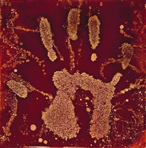
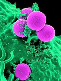
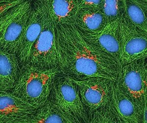
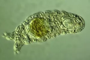
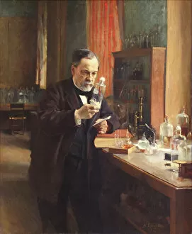
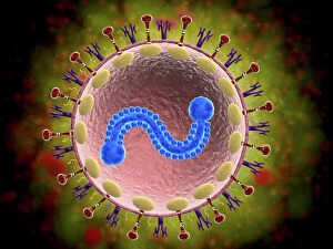
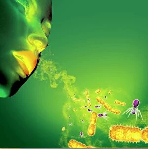
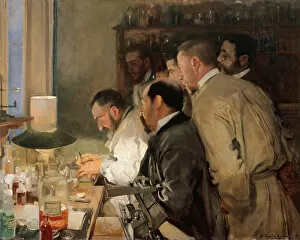
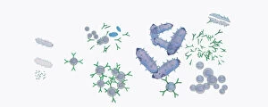
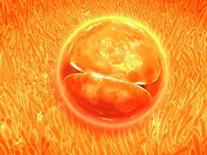
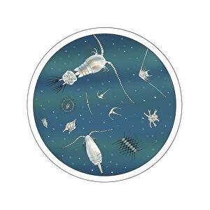
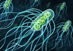
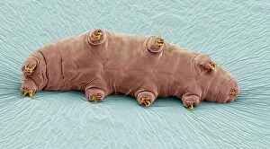
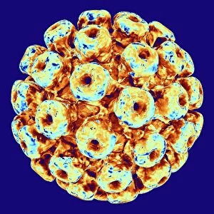
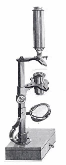
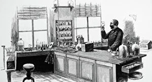
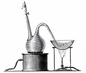
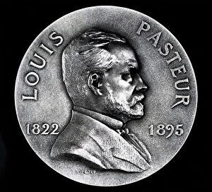
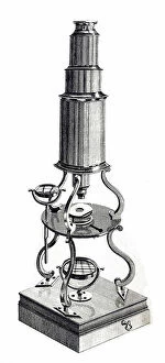
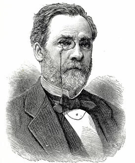
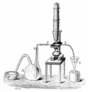
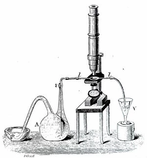
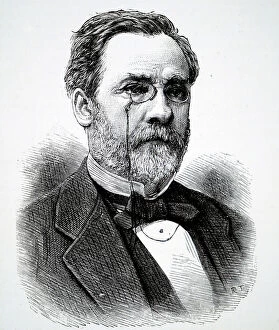
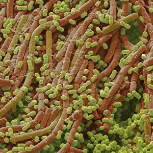
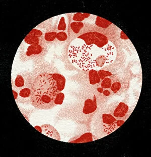
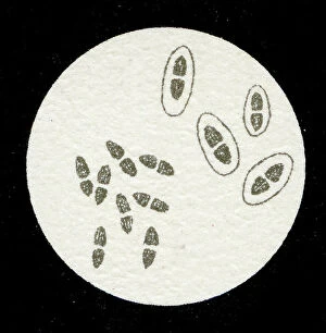
Microbiology, the fascinating world of tiny organisms and their impact on our lives. From the resilient Tardigrade or Water Bear that can survive extreme conditions to HeLa cells, a remarkable line of immortal human cells used in countless scientific studies. In Picture No. 11675590, we witness a mesmerizing light micrograph C017 / 8299 showcasing the intricate details of these microscopic wonders. The Neutrophil engulfing MRSA in SEM C018 / 8596 reminds us of the constant battle between our immune system and harmful bacteria. The beauty continues with another light micrograph C017 / 8298 capturing HeLa cells in all their glory. These cells have revolutionized medical research and paved the way for numerous breakthroughs. Yoghurt bacteria play a crucial role in transforming milk into this beloved creamy treat, highlighting how microbiology impacts even our daily diet. Stem cell research offers hope for regenerative medicine as we explore their incredible potential to repair and replace damaged tissues. Water bear once again graces us with its presence in light micrograph C016 / 8581, showcasing its unique anatomy under the microscope lens. We pay homage to Louis Pasteur, an influential figure who laid the foundation for modern microbiology through his groundbreaking discoveries and advancements in vaccination techniques. A glimpse into the invisible world reveals a microscopic view of human respiratory syncytial virus - a reminder that not all microbes are friendly companions. Embryo development captured just 24-36 hours after fertilization showcases nature's miracle unfolding before our eyes - each step meticulously orchestrated by molecular processes at work. And finally, artwork F008 /3245 portrays flu virus particles with artistic flair while reminding us of their ability to cause widespread illness if left unchecked. Microbiology unravels mysteries hidden from plain sight; it is both awe-inspiring and humbling as we delve deeper into this intricate realm where life's tiniest inhabitants shape our world.