
Membranelles Collection
Membranelles are fascinating structures found in various ciliate protozoans, such as Chilodonella and Climacostomum


All Professionally Made to Order for Quick Shipping
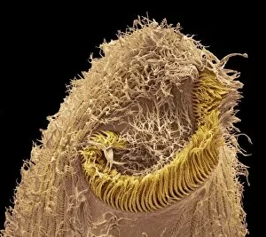
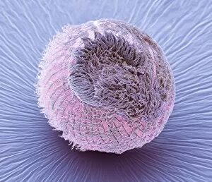
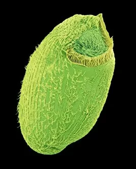
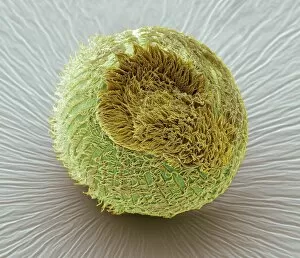
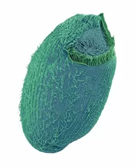
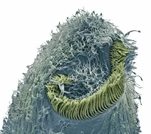
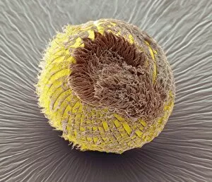
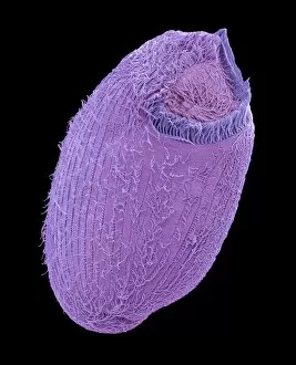
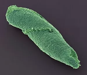
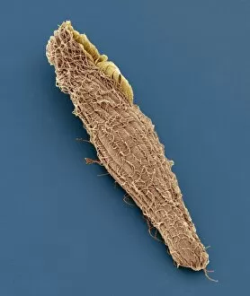
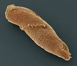
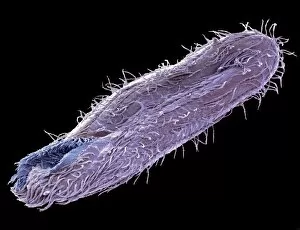
Membranelles are fascinating structures found in various ciliate protozoans, such as Chilodonella and Climacostomum. These tiny organisms have been captured in stunning detail using scanning electron microscopy (SEM), revealing the intricate beauty of their membranelles. In one SEM image, we observe a Chilodonella ciliate protozoan with its membranelles delicately arranged along its body. These hair-like structures serve multiple functions, including locomotion and feeding. They create rhythmic waves that propel the organism through water while also capturing food particles. Moving on to Climacostomum protozoans, SEM images showcase different species with distinct arrangements of membranelles. In one close-up shot, we witness the mesmerizing pattern formed by these structures on the surface of a Climacostomum specimen. The precise alignment suggests an evolved adaptation for efficient movement and prey capture. Another SEM image highlights Spirostomum ciliate protozoans exhibiting their unique membranelle arrangement. The spiraling bands of these hair-like projections create a striking visual effect as they undulate rhythmically during locomotion. Blepharisma ciliate protozoans also possess remarkable membranelles, as revealed in another captivating SEM image. Their elongated bodies bear rows of neatly organized hair-like appendages that aid in both swimming and feeding activities. These SEM images not only provide us with awe-inspiring visuals but also offer valuable insights into the diversity and complexity of these microorganisms' membrane systems. Membranelles play crucial roles in their survival by enabling them to navigate their aquatic habitats effectively while securing sustenance from surrounding resources. Studying these intricately designed membranelles helps scientists understand how structure relates to function at microscopic scales. Moreover, it highlights nature's incredible ability to adapt and evolve even within seemingly simple organisms like ciliates.