
Mandibles Collection (#3)
Mandibles: Nature's Mighty Jaws A panoramic dental X-ray reveals the intricate structure of human mandibles, crucial for chewing and speaking


All Professionally Made to Order for Quick Shipping
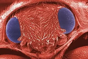
Mandibles: Nature's Mighty Jaws A panoramic dental X-ray reveals the intricate structure of human mandibles, crucial for chewing and speaking. The red-barbed ant, captured under a scanning electron microscope, showcases its formidable mandibles used for defense and capturing prey. Witness the trap-jaw ant with its menacing mandibles open wide, ready to snap shut on unsuspecting victims in the depths of Peru's Los Amigos Biological Station. In mid-flight, a red flour beetle displays its delicate yet powerful mandibles that aid in feeding and survival. Delve into history as we explore an artwork depicting the infamous Black Death rat flea, known for transmitting disease through bites using its sharp mandibles. An X-ray image reveals a fractured jawbone, highlighting the strength of our own jaws and their vulnerability to injury. Marvel at Darwin's beetle as it proudly flaunts its unique mandible adaptations that assist in courtship rituals and territorial disputes. Watch closely as a poppy bee skillfully cuts a piece of petal using its precise mandibles to create nesting material amidst Germany's blooming fields in June. The jumping spider from Orvieto demonstrates how it uses its specialized iridescent mandibles not only for hunting but also attracting mates during May's mating season in Italy. Behold the majesty of an army ant major soldier stationed at Parque da Onca Parda Private Reserve in Sao Paulo; armed with massive curved mandibles designed for battle and colony defense. The regal jumping spider captivates us with close-up shots showcasing vibrant iridescent colors on their impressive set of jaws ormandibless A cuckoo bee finds solace by clamping onto vegetation with her trusty pair ofmandibless after dawn breaks over Hertfordshire, England - truly nature’s ingenious adaptation for survival.