
Amygdala Collection
The amygdala, nestled in the medulla oblongata of the brain, is a captivating masterpiece of nature's design


All Professionally Made to Order for Quick Shipping
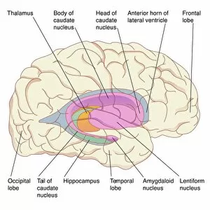
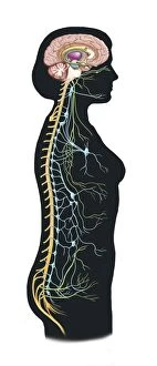
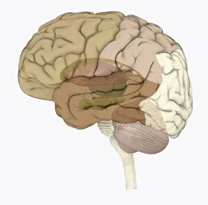
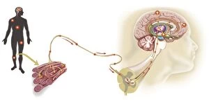
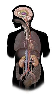
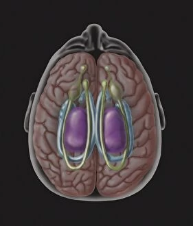
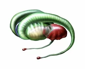
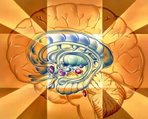
The amygdala, nestled in the medulla oblongata of the brain, is a captivating masterpiece of nature's design. Like an intricate artwork, it plays a crucial role in our emotions and memories. Adjacent to the basal ganglia, another artistic creation within our brains, the amygdala orchestrates the flow of information with finesse. A digital illustration vividly portrays this directionality, showcasing its significance in processing emotional stimuli. Within the human body's autonomic nervous system and limbic system lies this enigmatic structure - the amygdala. It forms an integral part of our brain's limbic system, influencing our responses to various situations. Highlighted alongside the striatum in a mesmerizing digital illustration within a human brain, we witness how these regions work together harmoniously. They shape our behaviors and decision-making processes as if they were brushstrokes on canvas. From childhood to adulthood, illustrations depicting profiles of heads reveal how the amygdala evolves over time. Its presence remains constant but matures along with us as we navigate life's complexities. Regardless of gender or age, every individual possesses their own unique version of this remarkable organ - showcased beautifully through digital illustrations capturing both female and male human brains. Intriguingly enough, specific areas within our brains are dedicated to facial recognition - one such area being closely intertwined with none other than the amygdala itself. A head profile reveals this connection like an artist revealing their inspiration behind a masterpiece. Delving deeper into cerebral architecture through yet another head profile illustration unveils not only motor cortexes but also frontal areas and auditory/visual cortices working hand-in-hand with that ever-present guardian: The Amygdala. As we marvel at these captivating visual representations highlighting different aspects of this extraordinary structure known as "amygdala, " let us appreciate its profound impact on shaping who we are as sentient beings – forever etched onto humanity's canvas.