Fraud Blocker Skip to main content

Alga Collection (#5)

"Discovering the Intricate Beauty of Algae: From Ernst Haeckel's Art to 19th-Century Cyanotypes and SEM Images" Immerse yourself in the captivating world of algae

Background imageAlga Collection: Algae cell wall, SEM

Algae cell wall, SEM
Algae cell wall. Coloured scanning electron micrograph (SEM) of cellulose microfibrils (strands) in a Chaetomorpha sp. algal cell wall

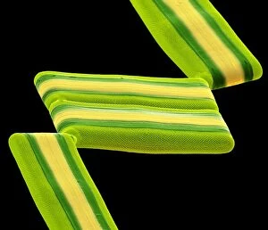
Background imageAlga Collection: Tabellaria diatoms, SEM C016 / 9599

Tabellaria diatoms, SEM C016 / 9599
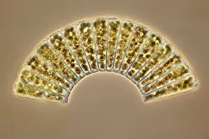
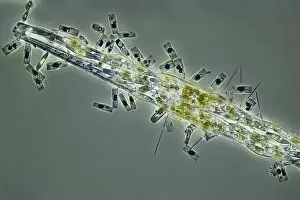
Tabellaria diatoms. Coloured scanning electron micrograph (SEM) of a Tabellaria sp. diatom colony. Diatoms are a group of photosynthetic, single-celled algae containing about 10, 000 species

Background imageAlga Collection: Coastal wildlife, artwork C016 / 7192

Coastal wildlife, artwork C016 / 7192
Coastal wildlife. Computer artwork showing the varying forms of wildlife found at different depths on a steep wave-swept Mediterranean coast

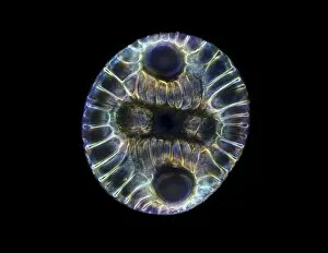
Background imageAlga Collection: Fossil diatom, light micrograph C016 / 8603

Fossil diatom, light micrograph C016 / 8603
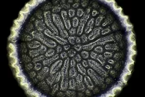
Fossil diatom. Darkfield illuminated light micrograph of a fossilised diatom. Diatoms are a group of photosynthetic, single-celled algae containing about 10, 000 species

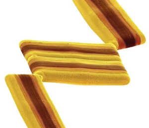
Background imageAlga Collection: Tabellaria diatoms, SEM C016 / 9600

Tabellaria diatoms, SEM C016 / 9600
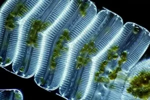
Tabellaria diatoms. Coloured scanning electron micrograph (SEM) of a Tabellaria sp. diatom colony. Diatoms are a group of photosynthetic, single-celled algae containing about 10, 000 species

Background imageAlga Collection: Tabellaria diatoms, SEM C016 / 9601

Tabellaria diatoms, SEM C016 / 9601
Tabellaria diatoms. Coloured scanning electron micrograph (SEM) of a Tabellaria sp. diatom colony. Diatoms are a group of photosynthetic, single-celled algae containing about 10, 000 species

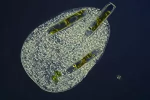
Background imageAlga Collection: Ciliate protozoan ingesting algae

Ciliate protozoan ingesting algae. Darkfield illuminated light micrograph of a ciliate protozoan ingesting green algae (green)

Background imageAlga Collection: Rodinia prehistoric landscape, artwork

Rodinia prehistoric landscape, artwork
Rodinia prehistoric landscape. Artwork showing a landscape at the time of the Rodinia supercontinent (1.1 billion to 750 million years ago) during the Neoproterozoic Era

Background imageAlga Collection: Haematococcus alga, light micrograph

Haematococcus alga, light micrograph
Haematococcus alga. Differential interference contrast (DIC) micrograph of a Haematococcus sp. green alga. Magnification: x670, when printed 10 centimetres wide

Background imageAlga Collection: Seaweed farming, Indonesia

Seaweed farming, Indonesia. Women processing harvested seaweed (red algae) at a small village in Takalar, Sulawesi, Indonesia. This seaweed will be used to make agar agar

Background imageAlga Collection: Microcoleus cyanobacteria, SEM

Microcoleus cyanobacteria, SEM
Microcoleus cyanobacteria. Coloured scanning electron micrograph (SEM) of Microcoleus sp. filamentous cyanobacteria (blue-green algae). Magnification: x 1300, when printed 10 centimetres wide

Background imageAlga Collection: Microalgae, light micrograph

Microalgae, light micrograph
Microalgae. Differential interference contrast (DIC) micrograph of Botryococcus braunii (round), and other species of green alga. Magnification: x278, when printed 10 centimetres wide

Background imageAlga Collection: Seaweed farming, Bali

Seaweed farming, Bali. Farmer working on his seaweed (red algae) farm off the coast of Kutuh, Bali, Indonesia. This seaweed will be used to make agar agar

Background imageAlga Collection: Haematococcus algae, light micrograph

Haematococcus algae, light micrograph
Haematococcus algae. Differential interference contrast (DIC) micrograph of a colony of Haematococcus sp. green alga. Magnification: x220, when printed 10 centimetres wide

Background imageAlga Collection: Ciliate protozoan, light micrograph

Ciliate protozoan, light micrograph
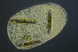
Ciliate protozoan. Darkfield illuminated light micrograph of a ciliate protozoan, showing green algae (green) that it has ingested

Background imageAlga Collection: Barents Sea algal bloom, satellite image

Barents Sea algal bloom, satellite image. North is at top, with land areas outlined in black. Algal blooms (green, top) have formed in the Barents Sea off northern Scandinavia (lower left)

Background imageAlga Collection: Dried seaweed specimens C016 / 5977

Dried seaweed specimens C016 / 5977
Dried seaweed (Chorda filum) specimens. Herbarium sheet from the collection held at the Natural History Museum, London, UK

Background imageAlga Collection: Intertidal zone, South Africa C017 / 1933

Intertidal zone, South Africa C017 / 1933
Intertidal zone. Time-exposure image of the sea lapping at algae-covered intertidal rocks. Photographed at Platbank, Goukamma Marine Protected Area, Western Cape, South Africa

Background imageAlga Collection: Surf fishing, South Africa C017 / 1934

Surf fishing, South Africa C017 / 1934
Surf fishing. Time-exposure image of a man fishing at dusk in the surf amongst algae-covered intertidal rocks. Photographed in the Goukamma Marine Protected Area, Western Cape, South Africa

Background imageAlga Collection: Scenedesmus algae, SEM C014 / 1462

Scenedesmus algae, SEM C014 / 1462
Scenedesmus algae. Coloured scanning electron micrograph (SEM) of Scenedesmus sp. algae. Scenedesmus is a nonmotile colonial green algae that has its cells arranged in a row

Background imageAlga Collection: Coccolithophore alga, SEM C019 / 0233

Coccolithophore alga, SEM C019 / 0233
Coccolithphore. Coloured scanning electron micrograph (SEM) of part of a coccolithophore unicellular marine alga. It is surrounded by a skeleton (coccosphere) of calcium carbonate plates (coccoliths)

Background imageAlga Collection: Coccolithophore alga, SEM C019 / 0234

Coccolithophore alga, SEM C019 / 0234
Coccolithphore. Coloured scanning electron micrograph (SEM) of part of a coccolithophore unicellular marine alga. It is surrounded by a skeleton (coccosphere) of calcium carbonate plates (coccoliths)

Background imageAlga Collection: Coccolithophore alga, SEM C019 / 0224

Coccolithophore alga, SEM C019 / 0224
Coccolithphore. Coloured scanning electron micrograph (SEM) of a coccolithophore unicellular marine alga. It is surrounded by a skeleton (coccosphere) of calcium carbonate plates (coccoliths)

Background imageAlga Collection: Diatom frustules (SEM) C014 / 2412

Diatom frustules (SEM) C014 / 2412
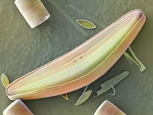
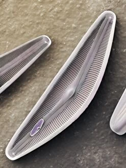
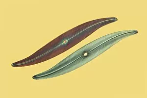
Diatom frustules. Coloured scanning electron micrograph of diatom frustules (Cymbella gastroides). Diatoms are a group of photosynthetic, single-celled algae containing about 10, 000 species

Background imageAlga Collection: Diatom frustules (SEM) C014 / 2147

Diatom frustules (SEM) C014 / 2147
Diatom frustules. Coloured scanning electron micrograph of the internal surface of a diatom frustule of Cymbella gastroides. The tiny diatom inside the cymbella is navicula sp

Background imageAlga Collection: Diatoms, light micrograph C014 / 4675

Diatoms, light micrograph C014 / 4675
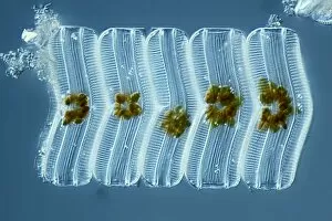
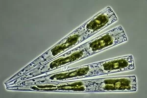
Diatoms. Differential interference contrast micrograph of a group of Meridion circulare diatoms. Diatoms are a group of photosynthetic, single-celled algae containing about 10, 000 species

Background imageAlga Collection: Diatom, light micrograph C014 / 4677

Diatom, light micrograph C014 / 4677
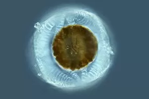
Diatom. Differential interference contrast micrograph of a Campylodiscus sp. diatom. Diatoms are a group of photosynthetic, single-celled algae containing about 10, 000 species

Background imageAlga Collection: Green algae, light micrograph C014 / 4670

Green algae, light micrograph C014 / 4670
Green algae. Differential interference contrast micrograph of a single green alga, showing the chloroplast (green) inside. Algae use the chloroplasts to produce food through photosynthesis

Background imageAlga Collection: Diatoms, light micrograph C014 / 4671

Diatoms, light micrograph C014 / 4671
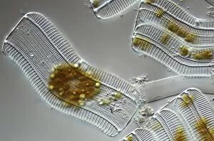
Diatoms. Differential interference contrast micrograph of a group of Achnantes longipes diatoms. Diatoms are a group of photosynthetic, single-celled algae containing about 10, 000 species

Background imageAlga Collection: Anabaena algae, SEM

Anabaena algae, SEM
Anabaena algae. Coloured scanning electron micrograph (SEM) of Anabaena sp. freshwater alga from a pond. Anabaena is a filamentous species of blue-green algae (cyanobacteria)

Background imageAlga Collection: Diatom, light micrograph C014 / 4669

Diatom, light micrograph C014 / 4669
Diatom. Differential interference contrast micrograph of an Achnantes longipes diatom. Diatoms are a group of photosynthetic, single-celled algae containing about 10, 000 species

Background imageAlga Collection: Diatoms, light micrograph C014 / 4668

Diatoms, light micrograph C014 / 4668
Diatoms. Differential interference contrast micrograph of a group of Achnantes longipes diatoms. Diatoms are a group of photosynthetic, single-celled algae containing about 10, 000 species

Background imageAlga Collection: Diatoms, light micrograph C014 / 4674

Diatoms, light micrograph C014 / 4674
Diatoms. Differential interference contrast micrograph of a group of Achnantes longipes diatoms. Diatoms are a group of photosynthetic, single-celled algae containing about 10, 000 species

Background imageAlga Collection: Arctic rocks and lichen

Arctic rocks and lichen (orange and grey). Lichens are formed from a symbiotic association between a fungus and an alga. The alga is contained within the fungus

Background imageAlga Collection: Ciliate protozoan, light micrograph C014 / 4676

Ciliate protozoan, light micrograph C014 / 4676
Ciliate protozoan. Differential interference contrast micrograph of a ciliate protozoan, showing the symbiotic green algae (Zoochlorellae, green) contained in vacuoles within

Background imageAlga Collection: Lichen, SEM C014 / 4736

Lichen, SEM C014 / 4736
Lichen. Coloured scanning electron micrograph (SEM) showing the structure of a lichen. Lichens are the result of a symbiotic relationship between a fungus and an alga

Background imageAlga Collection: Ciliate protozoan, light micrograph C014 / 4667

Ciliate protozoan, light micrograph C014 / 4667
Ciliate protozoan. Differential interference contrast micrograph of a ciliate protozoan, showing the symbiotic green algae (Zoochlorellae, green) contained in vacuoles within

Background imageAlga Collection: Plant evolutionary history, artwork

Plant evolutionary history, artwork
Plant evolutionary history. Artwork showing the stages in the evolution of plants during the history of the Earth. Around 1200 million years ago the Earths land areas were barren

Background imageAlga Collection: LM of the red algae, Plumaria elegans C014 / 8486

LM of the red algae, Plumaria elegans C014 / 8486
Plumaria algae. Light micrograph of part of the finely branched thallus of the red algae Plumaria elegans (family: Florideophycidae)

Background imageAlga Collection: LM of the red algae, Plumaria elegans C014 / 8488

LM of the red algae, Plumaria elegans C014 / 8488
Plumaria algae. Light micrograph of part of the finely branched thallus of the red algae Plumaria elegans (family: Florideophycidae)

Background imageAlga Collection: LM of the red algae, Plumaria elegans C014 / 8487

LM of the red algae, Plumaria elegans C014 / 8487
Plumaria algae. Light micrograph of part of the finely branched thallus of the red algae Plumaria elegans (family: Florideophycidae)

Background imageAlga Collection: Diatom, light micrograph C016 / 8606

Diatom, light micrograph C016 / 8606
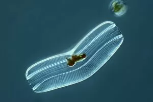
Diatom. Phase-contrast light micrograph of a diatom from the Mediterranean. Diatoms are a group of photosynthetic, single-celled algae containing about 10, 000 species

Background imageAlga Collection: Diatoms, light micrograph C016 / 8605

Diatoms, light micrograph C016 / 8605
Diatoms, Shearing contrast light micrograph. Diatoms are a group of photosynthetic, single-celled algae containing about 10, 000 species

Background imageAlga Collection: Diatoms, light micrograph C016 / 8598

Diatoms, light micrograph C016 / 8598
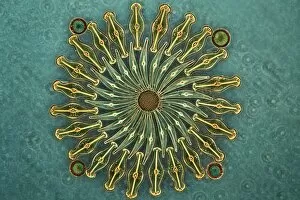
Diatoms. Interphase contrast light micrograph of a selection of differently shaped diatoms. Diatoms are a group of photosynthetic, single-celled algae containing about 10, 000 species

Background imageAlga Collection: Diatoms, light micrograph C016 / 8602

Diatoms, light micrograph C016 / 8602
Diatoms. Phase-contrast light micrograph of diatoms from the Mediterranean. Diatoms are a group of photosynthetic, single-celled algae containing about 10, 000 species

Background imageAlga Collection: Diatoms, light micrograph C016 / 8600

Diatoms, light micrograph C016 / 8600
Diatoms. Differential interference contrast (DIC) light micrograph of diatoms from the Mediterranean. Diatoms are a group of photosynthetic, single-celled algae containing about 10, 000 species

Background imageAlga Collection: Green algae, light micrograph C016 / 8595

Green algae, light micrograph C016 / 8595
Green algae, Interphase contrast light micrograph. Magnification: x280, when printed 10 centimetres wide

Background imageAlga Collection: Fossil diatom, light micrograph C016 / 8586

Fossil diatom, light micrograph C016 / 8586
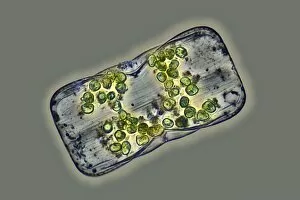
Fossil diatom. Interphase contrast light micrograph of a fossilised diatom. Diatoms are a group of photosynthetic, single-celled algae containing about 10, 000 species


All Professionally Made to Order for Quick Shipping

"Discovering the Intricate Beauty of Algae: From Ernst Haeckel's Art to 19th-Century Cyanotypes and SEM Images" Immerse yourself in the captivating world of algae, as we delve into its mesmerizing artistry and scientific wonders. Ernst Haeckel, a renowned biologist and artist, showcases the delicate intricacy of diatom algae through his stunning illustrations. These masterpieces reveal the symmetrical beauty hidden within these microscopic organisms. Step back in time with pressed seaweed specimens C016 / 6127 from centuries ago. Preserved meticulously, they offer a glimpse into the diverse forms and colors that algae can take on. The cyanotype technique used during the 19th century captures their essence in ethereal blue hues, adding an enchanting touch to their already fascinating nature. Calcareous phytoplankton observed under scanning electron microscopy (SEM) unravels another layer of algal marvels. Discosphaera tubifera, a coccolithophore species adorned with intricate calcium carbonate plates, showcases nature's architectural genius at work. Dictyota dichotoma emerges as a striking example of brown algae thriving along coastlines worldwide. Its branching structure creates an artistic spectacle underwater while providing essential habitats for marine life. Diatoms continue to amaze us with their geometric patterns when viewed under SEM imaging techniques. Their microscopic beauty is unveiled through meticulous detailing that reveals their remarkable diversity and ecological significance. Venturing further into aquatic realms, Fucus bulbosus or kelp stands tall as one of nature's giants beneath ocean waves. This majestic alga plays a crucial role in coastal ecosystems by offering shelter to numerous marine organisms. Pond life surprises us with its vibrant assortment of microorganisms where diatoms once again steal the show under SEM imaging techniques. Their intricate designs resemble miniature works of art floating amidst water bodies' tranquility.