Fraud Blocker Skip to main content

Urethra Collection (#4)

"Exploring the Intricacies of the Urethra

Background imageUrethra Collection: Voiding cystourethrogram, X-rays C018 / 0577

Voiding cystourethrogram, X-rays C018 / 0577
Voiding cystourethrogram. X-ray of a 54 year old male patient undergoing a voiding cystourethrogram. This procedure involves liquid containing a radio-opaque dye being placed in the bladder via a

Background imageUrethra Collection: Voiding cystourethrogram, X-rays C018 / 0578

Voiding cystourethrogram, X-rays C018 / 0578
Voiding cystourethrogram. Coloured X-ray of a 54 year old male patient undergoing a voiding cystourethrogram. This procedure involves liquid containing a radio-opaque dye being placed in the bladder

Background imageUrethra Collection: Voiding cystourethrogram, X-rays C018 / 0579

Voiding cystourethrogram, X-rays C018 / 0579
Voiding cystourethrogram. Coloured X-ray of a 54 year old male patient undergoing a voiding cystourethrogram. This procedure involves liquid containing a radio-opaque dye being placed in the bladder

Background imageUrethra Collection: Voiding cystourethrogram, X-rays C018 / 0465

Voiding cystourethrogram, X-rays C018 / 0465
Voiding cystourethrogram. Three coloured X-rays showing a 65 year old patient undergoing a voiding cystourethrogram. This procedure involves liquid containing a radio-opaque dye being placed in

Background imageUrethra Collection: Bladder stones, artwork

Bladder stones, artwork
Bladder stones. Computer artwork showing a section through a human bladder containing bladder stones (whiteish, centre). The urethra can be seen (vertical, lower centre)

Background imageUrethra Collection: Urinary system, artwork C015 / 6455

Urinary system, artwork C015 / 6455
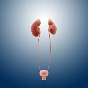
Urinary system. Computer artwork showing the organs of the human urinary system. The kidneys (red, top left and right) filter waste products such as urea from the blood (red and blue) to form urine

Background imageUrethra Collection: Urinary system, artwork C015 / 6454

Urinary system, artwork C015 / 6454
Urinary system. Computer artwork showing the organs of the human urinary system. The kidneys (red, top left and right) filter waste products such as urea from the blood to form urine

Background imageUrethra Collection: Male anatomy, artwork C015 / 6448

Male anatomy, artwork C015 / 6448
Male anatomy. Computer artwork showing the major organs of the human torso. From top to bottom: oesophagus (white, top centre), trachea (windpipe, pink, top centre)

Background imageUrethra Collection: Female anatomy, artwork C015 / 6447

Female anatomy, artwork C015 / 6447
Female anatomy. Computer artwork showing the major organs of the femaletorso. From top to bottom: oesophagus (white, top centre), trachea (windpipe, pink, top centre)

Background imageUrethra Collection: Single-kidney anatomy, artwork C016 / 2902

Single-kidney anatomy, artwork C016 / 2902
Single-kidney anatomy. Artwork showing an anterior (frontal) view of human kidneys in relation to the main torso blood vessels (vena cava, blue; aorta, red) and the male abdomen

Background imageUrethra Collection: Single-kidney anatomy, artwork C016 / 2901

Single-kidney anatomy, artwork C016 / 2901
Single-kidney anatomy. Artwork showing an anterior (frontal) view of human kidneys in relation to the main torso blood vessels (vena cava, blue; aorta, red) and bones of the male skeleton

Background imageUrethra Collection: Single-kidney anatomy, artwork C016 / 2900

Single-kidney anatomy, artwork C016 / 2900
Single-kidney anatomy. Artwork showing an anterior (frontal) view of human kidneys in relation to the main torso blood vessels (vena cava, blue; aorta, red) and bones of the skeleton

Background imageUrethra Collection: Single-kidney anatomy, artwork C016 / 2896

Single-kidney anatomy, artwork C016 / 2896
Single-kidney anatomy. Artwork showing an anterior (frontal) view of human kidneys in relation to the main torso blood vessels (vena cava, blue; aorta, red)

Background imageUrethra Collection: Urinary system, artwork C013 / 4673

Urinary system, artwork C013 / 4673
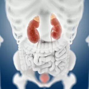
Urinary system. Computer artwork showing an anterior view of the kidneys, the main organs of the urinary system, relative to the skeleton and abdominal organs (white)

Background imageUrethra Collection: Urinary system, artwork C013 / 4670

Urinary system, artwork C013 / 4670
Urinary system. Computer artwork showing the kidneys (top), the main organs of the urinary system. The kidneys are located in the upper abdomen

Background imageUrethra Collection: Urinary system, artwork C013 / 4669

Urinary system, artwork C013 / 4669
Urinary system. Computer artwork showing the kidneys (top), the main organs of the urinary system. The kidneys are located in the upper abdomen

Background imageUrethra Collection: Interstitial cystitis

Interstitial cystitis. Conceptual image of a naked woman with an inflamed bladder (lower centre) caused by interstitial cystitis

Background imageUrethra Collection: Human urinary system, artwork

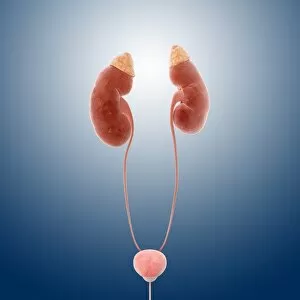
Human urinary system, artwork
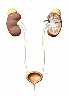
Urinary system. Computer artwork of a male torso and the urinary system, showing kidney, urethra, bladder and suprarenal gland

Background imageUrethra Collection: Healthy male bladder, artwork

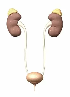
Healthy male bladder, artwork

Background imageUrethra Collection: Healthy urinary system, artwork

Healthy urinary system, artwork

Background imageUrethra Collection: Incontinence control, conceptual image

Incontinence control, conceptual image. Computer artwork of G-clamps squeezing a urethra

Background imageUrethra Collection: Urethral stricture, artwork

Urethral stricture, artwork
Urethral stricture. Computer artwork of a narrowing of the urethra. This can be caused by injury or disease, such as urinary tract infections or other causes of urethritis

Background imageUrethra Collection: Human internal organs, diagram

Human internal organs, diagram
Human internal organs. Diagram showing the human internal organs in the head and torso. In the head, the organs and cavities shown are: the brain (encephalon), the cerebellum (part of the brain)

Background imageUrethra Collection: Human internal organs, artwork

Human internal organs, artwork
Human internal organs. Artwork showing the human internal organs in the head and torso. In the head, the organs and cavities shown are: the brain (encephalon), the cerebellum (part of the brain)

Background imageUrethra Collection: Prostate gland anatomy, artwork

Prostate gland anatomy, artwork
Artwork of the anatomy of the prostate gland showing three glandular regions: peripheral zone (PZ) making up 70% of the gland and surrounds the urethra; central zone (CZ)

Background imageUrethra Collection: Bladder anatomy, artwork

Bladder anatomy, artwork

Background imageUrethra Collection: Urinary system, artwork

Urinary system, artwork
Urinary system. Artwork showing the human urinary system on an outline of the human body, showing the location of the kidneys (upper left and upper right) and bladder (lower centre)

Background imageUrethra Collection: Bladder expansion, artwork

Bladder expansion, artwork
Bladder expansion. Sequential artwork showing the external structure of an expanding bladder. Successive shades of lighter pink show the bladder expanding greatly in volume from its resting state


All Professionally Made to Order for Quick Shipping

"Exploring the Intricacies of the Urethra: A Fascinating Journey Through Female Bladder Artwork" Delve into the captivating world of the urethra as we embark on a visual exploration through remarkable artwork. Witness an exquisite depiction of a section through the female bladder, revealing its intricate structure and function. Intriguingly, our journey takes us further as we encounter a lithograph showcasing a colony of Micrococcus Gonorrhoea found in the urethra back in 1906. This historical representation sheds light on early scientific discoveries surrounding this microorganism. Moving forward, we delve into cross-sectional anatomy with rectal exams conducted on normal males. These illustrations provide valuable insights into male reproductive systems and urinary tracts using both traditional and ultrasound technologies. Marvel at digital cross-section illustrations that unravel the complexity of male reproductive systems, offering an unprecedented glimpse into their inner workings. Additionally, microscopic views reveal Neisseria gonorrhoeae—a bacterium responsible for causing infections within this delicate organ. Surprisingly, our exploration extends beyond human anatomy to include intriguing aspects such as Vandellia cirrhosa or candiru—an infamous fish known for its peculiar parasitic behavior within urethras. Such unique encounters remind us of nature's diversity and adaptability. Lastly, let us not forget about kidney health; witness an early stage kidney cancer with a visible tumor on this vital organ. This serves as a reminder to prioritize regular check-ups and maintain awareness regarding potential risks associated with urological health. Through these captivating visuals encompassing both artistry and medical science, we gain profound appreciation for the intricacies housed within our urinary systems—highlighting their significance in maintaining overall well-being.