Fraud Blocker Skip to main content

Replicating Collection (#2)

"Unveiling the Art of Replication: From Monks Copying Manuscripts to Budding HIV Particles" In the ancient halls of monasteries

Background imageReplicating Collection: DNA replication by helicase enzyme C013 / 9382

DNA replication by helicase enzyme C013 / 9382
Computer artwork of DNA Helicase breaking apart the hydrogen bonds of a DNA strand for replication. Helicases are a class of enzymes vital to all living organisms

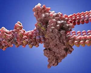
Background imageReplicating Collection: DNA polymerase molecule C013 / 7909

DNA polymerase molecule C013 / 7909
DNA polymerase. Molecular model of a molecule of DNA polymerase (blue) replicating a strand of DNA (deoxyribonucleic acid, pink and turquoise). The secondary structure of the DNA polymerase is shown

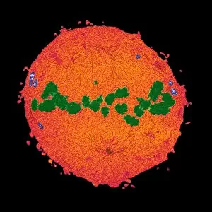
Background imageReplicating Collection: Mitosis, TEM

Mitosis, TEM
Mitosis, coloured transmission electron micrograph (TEM). Longitudinal section through a human T cell in metaphase. During mitosis two daughter nuclei are formed from one parent nucleus

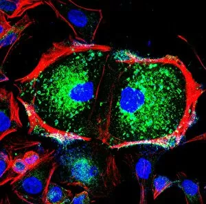
Background imageReplicating Collection: Dividing cell, light micrograph

Dividing cell, light micrograph
Cell division. Fluorescent light micrograph of a cell that has divided by mitosis, the asexual replication of a cell into two new cells

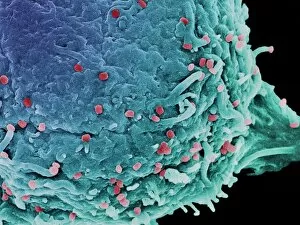
Background imageReplicating Collection: Vaccinia virus particles, SEM

Vaccinia virus particles, SEM
Vaccinia virus particles. Coloured scanning electron micrograph (SEM) of vaccinia virus particles (red) budding from a cell (blue and green) after replicating

Background imageReplicating Collection: Viruses attacking a cells DNA

Viruses attacking a cells DNA, conceptual computer artwork. Some viruses replicate themselves by inserting their own DNA (deoxyribonucleic acid) into that of a host cell

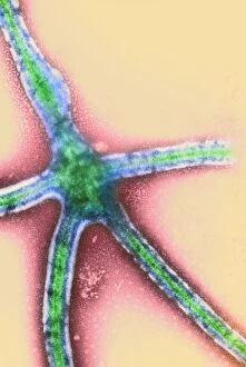
Background imageReplicating Collection: Ebola virus replication, TEM

Ebola virus replication, TEM

Background imageReplicating Collection: Dengue fever virus replication, TEM

Dengue fever virus replication, TEM

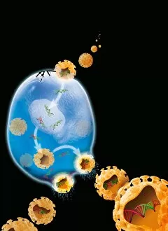
Background imageReplicating Collection: Virus replication cycle, artwork

Virus replication cycle, artwork. The cycle starts at upper centre, as virus particles (orange) enter a cell (blue). Once in the cell the viral genetic material, deoxyribonucleic acid (DNA, green)

Background imageReplicating Collection: Paramyxovirus, TEM

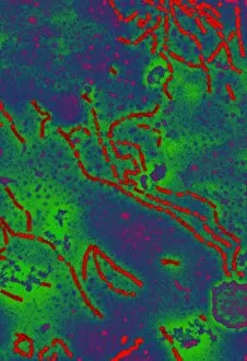
Paramyxovirus, TEM
Paramyxovirus. Transmission electron micrograph (TEM) of paramyxovirus ribonuclear protein helices (red strands). These helices contain the viral genetic material, ribonucleic acid (RNA)

Background imageReplicating Collection: Simian AIDS viruses, TEM

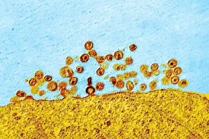
Simian AIDS viruses, TEM
Simian AIDS virus particles, coloured transmission electron micrograph (TEM). These virus particles (small, round) are bursting out of a cell (across bottom) after using the cell to replicate

Background imageReplicating Collection: West Nile virus, TEM

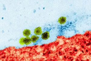
West Nile virus, TEM
West Nile virus particles. Coloured transmission electron micrograph (TEM) of West Nile virus (WNV) particles (green) next to a host cell (red)

Background imageReplicating Collection: Herpes simplex virus infection, TEM

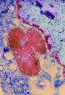
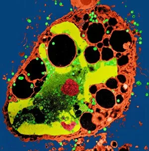
Herpes simplex virus infection, TEM
Herpes simplex virus infection. Coloured transmission electron micrograph (TEM) of a section through a cell being destroyed by herpes simplex virus infection

Background imageReplicating Collection: Mitosis, artwork

Mitosis, artwork

Background imageReplicating Collection: Dividing bacteria, artwork

Dividing bacteria, artwork

Background imageReplicating Collection: Taq polymerase replicating DNA

Taq polymerase replicating DNA, molecular model. The Taq polymerase is blue, the two strands of DNA are green, and the nucleotide bases are grey (carbon), pink (nitrogen)

Background imageReplicating Collection: Protocell proliferation, artwork

Protocell proliferation, artwork
Protocell proliferation. Image 5 of 5. Artwork showing a protocell (artificial cell) dividing to produce two daughter cells

Background imageReplicating Collection: Viral RNA replication cycle, artwork

Viral RNA replication cycle, artwork
Viral RNA replication cycle. Computer artwork showing the three stages of the replication cycle that forms double-stranded DNA (deoxyribonucleic acid)

Background imageReplicating Collection: Energised protocell, artwork

Energised protocell, artwork
Energised protocell. Image 4 of 5. Artwork showing light photons energising a protocell (artificial cell). This protocell was formed from a synthetic primeval soup containing PNA

Background imageReplicating Collection: Influenza viruses, TEM

Influenza viruses, TEM
Influenza viruses. Coloured transmission electron micrograph (TEM) of influenza (flu) viruses (yellow) budding from a host cell. The host cell nucleus is black

Background imageReplicating Collection: Retrovirus genome replication, artwork

Retrovirus genome replication, artwork
Retrovirus genome replication. Computer artwork showing the replication and expression of retroviral RNA (ribonucleic acid)

Background imageReplicating Collection: Enterococcus faecalis dividing, TEM

Enterococcus faecalis dividing, TEM
HPA Colindale 12/01/12 12:30:37 15000 7.0 80.0 Imaging Enterococcus faecalis 1.5 AmMob plus wash -170.96 657.83 . XpixCal=1.242 YpixCal=1.242 Unit=nm ##fv3

Background imageReplicating Collection: Dividing bacteria, computer artwork

Dividing bacteria, computer artwork
Bacteria dividing, computer artwork

Background imageReplicating Collection: Cell division, artwork

Cell division, artwork
Cell division. Computer artwork of an animal cell undergoing mitosis (nuclear division) and cytokinesis (cell division) to form two identical daughter cells (bottom)

Background imageReplicating Collection: Bacteria with integrated foreign bacteria

Bacteria with integrated foreign bacteria. Computer artwork showing a section of foreign DNA (deoxyribonucleic acid, blue) integrated into a bacterial chromosome (green)

Background imageReplicating Collection: HIV replication cycle, diagram

HIV replication cycle, diagram
HIV replication cycle. Diagram and magnified views showing the retroviral process by which the human immunodeficiency virus (HIV) replicates

Background imageReplicating Collection: Genetic molecular mechanisms, artwork

Genetic molecular mechanisms, artwork

Background imageReplicating Collection: Bacterial DNA, conceptual artwork

Bacterial DNA, conceptual artwork
Bacterial DNA, conceptual computer artwork. Bacterial cells containing two molecules of DNA (deoxyribonucleic acid, represented by letters)

Background imageReplicating Collection: DNA replication fork, artwork

DNA replication fork, artwork
DNA replication fork. Diagram showing the cyclic sequence (right) for replication of DNA (deoxyribonucleic acid). Details of the DNA are at left

Background imageReplicating Collection: Nanobot replication

Nanobot replication. Self-contained nanobots replicating in a factory. Nanotechnology involves the use of current and future technology to build microscopically small probes

Background imageReplicating Collection: DNA replication process, diagram

DNA replication process, diagram
DNA replication process. Diagram showing various stages in the replication of DNA (deoxyribonucleic acid). The process starts (top) when initiator proteins (blue-grey) separate the strands of DNA

Background imageReplicating Collection: Bacterial replication, artwork

Bacterial replication, artwork
Bacterial replication. Computer artwork showing a single bacterium dividing to form two identical daughter cells. This process is a form of asexual reproduction and is known as binary fission

Background imageReplicating Collection: DNA replication

DNA replication. Computer artwork of a DNA (deoxyribonucleic acid) molecule replicating. DNA is composed of two strands twisted into a double helix (top)

Background imageReplicating Collection: DNA replication, artwork

DNA replication, artwork
DNA replication. Computer artwork of a DNA (deoxyribonucleic acid) molecule replicating. DNA is composed of two strands twisted into a double helix


All Professionally Made to Order for Quick Shipping

"Unveiling the Art of Replication: From Monks Copying Manuscripts to Budding HIV Particles" In the ancient halls of monasteries, dedicated monks meticulously replicate manuscripts, preserving knowledge for generations to come. (Monks Copying Manuscript) Underneath our very noses, a microscopic world thrives with replication as Candida fungus multiplies and spreads its presence. (Candida fungus, SEM) Witness the mesmerizing dance of yeast cells dividing, their intricate process captured in stunning detail under the scanning electron microscope. (Dividing yeast cells, SEM) The relentless nature of life continues as herpes virus replicates within host cells, perpetuating its existence and causing discomfort. (Herpes virus replicating) Delving deeper into unseen realms reveals intestinal protozoan parasites undergoing replication - an astonishing sight through the transmission electron microscope. (Intestinal protozoan parasites, TEM) Behold the striking image of Vesicular stomatitis virus particles multiplying relentlessly inside host cells - a testament to their survival instincts. (Vesicular stomatitis virus, TEM) Peering into the microcosm once again uncovers E. coli bacterium replicating with remarkable precision and efficiency - an awe-inspiring feat captured by the transmission electron microscope. (E. coli bacterium, TEM) Salmonella bacterium divides before our eyes in this captivating scanning electron microscopy image – showcasing how they propagate their lineage through division. (Salmonella bacterium dividing, SEM) Cell division unfolds like a delicate ballet on a grand stage; witness this breathtaking spectacle through high-resolution scanning electron microscopy imagery. (Cell division, SEM) Plant cell mitosis comes alive under light micrography's gentle touch – revealing nature's elegant way of creating new life from existing ones. (Plant cell mitosis, light micrograph) Budding HIV particles emerge from infected cells like tiny alien spacecrafts ready to infect anew - a hauntingly beautiful sight captured by scanning electron microscopy.