Fraud Blocker Skip to main content

Microbiological Collection

Microbiological wonders unfold in this captivating collection of images

Background imageMicrobiological Collection: Budding yeast cell

Budding yeast cell. Computer artwork of asection through a yeast ( Candida albicans )cell that is reproducing asexually. A daughtercell (top left) is budding from the parent cell

Background imageMicrobiological Collection: Avian flu virus

Avian flu virus, computer artwork. A virus is a tiny pathogenic particle comprising genetic material enclosed in a protein coat. The coat contains surface proteins (spikes)

Background imageMicrobiological Collection: Calcareous phytoplankton, SEM

Calcareous phytoplankton, SEM
Calcareous phytoplankton. Coloured scanning electron micrograph (SEM) of the external mineralised structures (coccospheres) of small marine algal organisms called coccolithophores

Background imageMicrobiological Collection: Neutrophil engulfing MRSA, SEM C018 / 8596

Neutrophil engulfing MRSA, SEM C018 / 8596
Neutrophil engulfing MRSA. Coloured scanning electron micrograph (SEM) of a neutrophil white blood cell (green) engulfing methicillin-resistant Staphylococcus aureus bacteria (MRSA, pink)

Background imageMicrobiological Collection: Acrosphaera radiolarian, SEM

Acrosphaera radiolarian, SEM
Acrosphaera radiolarian. Coloured scanning electron micrograph (SEM) of the shell of a Acrosphaera sp. radiolarian. Radiolaria are single-celled protozoans that are found in marine plankton

Background imageMicrobiological Collection: Anthrax cultures, historical diagram

Anthrax cultures, historical diagram
Anthrax cultures. 1876 diagram of Bacillus anthracis (Anthrax) bacteria cultured by Robert Koch. Along with Louis Pasteur, Koch is considered the founder of modern medical bacteriology

Background imageMicrobiological Collection: Diatom, SEM

Diatom, SEM
Diatom. Coloured scanning electron micrograph (SEM) of the Arachnoidiscus sp. diatom. The diatoms are a group of photosynthetic, single- celled algae containing about 10, 000 species

Background imageMicrobiological Collection: Diatom, SEM

Diatom, SEM
Diatom. Coloured scanning electron micrograph (SEM) of a Stephanopyxis sp. diatom. Diatoms are single-celled photosynthetic algae, of which there are about 100, 000 species

Background imageMicrobiological Collection: E. coli bacteria, SEM

E. coli bacteria, SEM
E. coli bacteria. Coloured scanning electron micrograph (SEM) of Escherichia coli bacteria. These bacteria are a normal part of the intestinal flora in humans and other animals

Background imageMicrobiological Collection: Diatom, SEM

Diatom, SEM
Diatom. Coloured scanning electron micrograph (SEM) of a Triceratium morlandii diatom. Diatoms are single-celled photosynthetic algae, of which there are about 100, 000 species

Background imageMicrobiological Collection: Herpes virus replicating

Herpes virus replicating, computer artwork.Viruses are only able to replicate in a host cell.The glycoprotein spikes in the virus envelope(green)

Background imageMicrobiological Collection: Salmonella bacteria, SEM

Salmonella bacteria, SEM
Salmonella bacteria, coloured scanning electron micrograph (SEM). These Gram-negative rod-shaped bacteria can cause food poisoning when eaten in contaminated food

Background imageMicrobiological Collection: Candida fungus, SEM

Candida fungus, SEM
Candida fungus. Coloured scanning electron micrograph (SEM) of budding threads (hyphae) of a Candida fungus. The budding areas are where asexual reproduction is producing new fungus cells

Background imageMicrobiological Collection: HIV particle

HIV particle, computer artwork. HIV (human immunodeficiency virus) causes AIDS (acquired immune deficiency syndrome). The virus consists of an RNA genome

Background imageMicrobiological Collection: Avian flu virus

Avian flu virus, computer artwork. A virus is a tiny pathogenic particle comprising genetic material enclosed in a protein coat. The coat contains surface proteins (spikes)

Background imageMicrobiological Collection: Cell types, artwork

Cell types, artwork
Cell types. Cutaway artwork with a scale bar (upper right, in micrometres) showing the relative sizes of eukaryotic cells (those containing a nucleus) and prokaryotic cells (those lacking a nucleus)

Background imageMicrobiological Collection: Norovirus particles, TEM

Norovirus particles, TEM
Norovirus particles. Coloured transmission electron micrograph (TEM) of norovirus particles. Norovirus is a genus of RNA (ribonucleic acid)

Background imageMicrobiological Collection: Tuberculosis bacteria

Tuberculosis bacteria. Computer artwork of Mycobacterium tuberculosis bacteria. These Gram- positive rod-shaped bacteria cause the disease tuberculosis

Background imageMicrobiological Collection: Pseudomonas aeruginosa bacteria, SEM

Pseudomonas aeruginosa bacteria, SEM
Pseudomonas aeruginosa bacteria, coloured scanning electron micrograph (SEM). These Gram-negative rod-shaped bacteria are found in soil and water, and as normal flora in the human intestine

Background imageMicrobiological Collection: Dividing yeast cells, SEM

Dividing yeast cells, SEM
Dividing yeast cells. Coloured scanning electron micrograph (SEM) of Schizosaccharomyces pombe yeast cells dividing. S. pombe is a single-celled fungus that is studied widely as a model organism for

Background imageMicrobiological Collection: Dividing yeast cells, SEM

Dividing yeast cells, SEM
Dividing yeast cells. Coloured scanning electron micrograph (SEM) of Schizosaccharomyces pombe yeast cells dividing. S. pombe is a single-celled fungus that is studied widely as a model organism for

Background imageMicrobiological Collection: Bacteria infecting a macrophage, SEM

Bacteria infecting a macrophage, SEM
Bacteria infecting a macrophage. Coloured scanning electron micrograph (SEM) of Mycobacterium tuberculosis bacteria (purple) infecting a macrophage white blood cell

Background imageMicrobiological Collection: Flagellate bacteria

Flagellate bacteria. Computer artwork of a rod- shaped bacteria with polar flagella. Bacteria with a morphology similar to this include Helicobacter pylori and Salmonella sp

Background imageMicrobiological Collection: E. coli bacteria, SEM

E. coli bacteria, SEM
E. coli bacteria. Coloured scanning electron micrograph (SEM) of Escherichia coli bacteria (purple) taken from the small intestine of a child. E

Background imageMicrobiological Collection: Salmonella bacteria, SEM

Salmonella bacteria, SEM
Salmonella bacteria, coloured scanning electron micrograph (SEM). Each rod-shaped structure is an individual bacterium. These bacteria occur mainly in human and animal intestines

Background imageMicrobiological Collection: Pseudomonas aeruginosa bacteria, SEM

Pseudomonas aeruginosa bacteria, SEM
Pseudomonas aeruginosa bacteria, coloured scanning electron micrograph (SEM). These Gram-negative rod-shaped bacteria are found in soil and water, and as normal flora in the human intestine

Background imageMicrobiological Collection: E. coli bacterium, TEM

E. coli bacterium, TEM
E. coli bacterium, coloured transmission electron micrograph (TEM). E. coli are Gram-negative rod-shaped bacteria that are part of the normal flora of the human gut

Background imageMicrobiological Collection: Water bear, light micrograph C016 / 8581

Water bear, light micrograph C016 / 8581
Water bear, differential interference contrast (DIC) light micrograph. Water bears, or tardigrades, (phylum Tardigrada) are small, water-dwelling

Background imageMicrobiological Collection: Louis Pasteur, French microbiologist

Louis Pasteur, French microbiologist
Louis Pasteur (1822-1895), French microbiologist and chemist. Louis Pasteur proved the germ theory of disease, which holds that germs attack the body from the outside

Background imageMicrobiological Collection: Macrophage engulfing TB bacteria, SEM

Macrophage engulfing TB bacteria, SEM
Macrophage engulfing TB bacteria. Coloured scanning electron micrograph (SEM) of a macrophage white blood cell (purple) engulfing a tuberculosis (Mycobacterium tuberculosis) bacterium (pink)

Background imageMicrobiological Collection: Infections spread by sneezing, artwork C013 / 5949

Infections spread by sneezing, artwork C013 / 5949
Infections spread by sneezing. Computer artwork showing virus particles (virions, blue) and bacteria (rod-shaped) in the spray produced by someone when they sneeze

Background imageMicrobiological Collection: Cup lichen (Cladonia floerkeana)

Cup lichen (Cladonia floerkeana)
Lichen fruiting bodies (red). This lichen species is Cladonia floerkeana, found on heaths and moorlands. Lichens are formed from a symbiotic association between a fungus and an alga

Background imageMicrobiological Collection: Pasteurs Jubilee celebrations, 1892

Pasteurs Jubilee celebrations, 1892
Pasteurs Jubilee celebrations. Artwork of British surgeon Joseph Lister (in gown, walking up steps, 1827-1912) and French chemist Louis Pasteur (being approached by Lister)

Background imageMicrobiological Collection: Diatom frustule, SEM

Diatom frustule, SEM
Diatom frustule. Coloured scanning electron micrograph (SEM) of the frustule of a Pinnularia sp. diatom. The diatoms are a group of photosynthetic, single-celled algae containing about 10

Background imageMicrobiological Collection: Flu virus particle, artwork F008 / 3245

Flu virus particle, artwork F008 / 3245
Flu virus particle, computer artwork. In the virus envelope are two types of protein spike, haemagglutinin (H, pink) and neuraminidase (N, orange), which determine the strain of virus

Background imageMicrobiological Collection: Plankton, artwork C016 / 3447

Plankton, artwork C016 / 3447
Plankton. Artwork of plankton as viewed under a magnifying glass

Background imageMicrobiological Collection: Salmonella bacteria, artwork C013 / 8818

Salmonella bacteria, artwork C013 / 8818
Salmonella bacteria, computer artwork. Salmonella sp. bacteria are gram-negative rod-shaped bacteria that have flagella (hair-like structures) that they use for locomotion

Background imageMicrobiological Collection: Foraminifera, SEM

Foraminifera, SEM
Foraminifera. Coloured scanning electron micrograph (SEM) of a selection of foraminiferans, including Oolina hexagona (bottom right) and a number of Lagena sp

Background imageMicrobiological Collection: Phagocytosis of fungal spores, SEM

Phagocytosis of fungal spores, SEM
Phagocytosis of a fungus spore. Coloured scanning electron micrograph (SEM) of a neutrophil (blue) phagocytosing (engulfing and destroying) spores from the fungus Aspergillus fumigatus (red)

Background imageMicrobiological Collection: Neutrophil cell trapping bacteria, SEM

Neutrophil cell trapping bacteria, SEM
Neutrophil cell trapping bacteria. Coloured scanning electron micrograph (SEM) of bacteria (rod-shaped) being trapped by a neutrophil cell

Background imageMicrobiological Collection: Influenza virus, computer artwork

Influenza virus, computer artwork
Influenza virus. Computer artwork of an influenza (flu) virus. The virus consists of a core of RNA (ribonucleic acid) genetic material surrounded by a protein coat

Background imageMicrobiological Collection: Microbiology caricature, 19th century

Microbiology caricature, 19th century
Microbiology caricature. The researcher is handling jars labelled as bacterial cultures, while the magnified view at left shows a caricatured appearance of microbes as seen under a microscope

Background imageMicrobiological Collection: Influenza virus particles, TEM

Influenza virus particles, TEM
Influenza virus particles, coloured transmission electron micrograph (TEM). This virus is the cause of the respiratory disease influenza (flu) in humans and other animals

Background imageMicrobiological Collection: Hepatitis C viruses, TEM

Hepatitis C viruses, TEM
Hepatitis C viruses. Coloured transmission electron micrograph (TEM) of hepatitis C virus particles (green) infecting cultured liver cells (purple)

Background imageMicrobiological Collection: Louis Pasteur, French microbiologist

Louis Pasteur, French microbiologist
Louis Pasteur (1822-1895), French microbiologist and chemist, in his laboratory. Louis Pasteur proved the germ theory of disease, which holds that germs attack the body from the outside

Background imageMicrobiological Collection: Dinoflagellate plankton, SEM

Dinoflagellate plankton, SEM
Dinoflagellate plankton. Coloured scanning electron micrograph (SEM) of the calcareous (calcium-based) test (shell) of a dinoflagellate Calciodinellum sp

Background imageMicrobiological Collection: Pseudomonas aeruginosa bacteria, SEM

Pseudomonas aeruginosa bacteria, SEM
Pseudomonas aeruginosa bacteria, coloured scanning electron micrograph (SEM). These Gram-negative rod-shaped bacteria are found in soil and water, and as normal flora in the human intestine

Background imageMicrobiological Collection: Water bear, SEM C016 / 9084

Water bear, SEM C016 / 9084
Water bear (Echiniscus granulatus). Coloured scanning electron micrograph (SEM) showing the underside of a water bear, or tardigrade (phylum Tardigrada)


All Professionally Made to Order for Quick Shipping

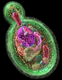
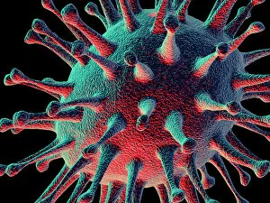
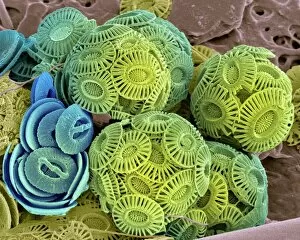
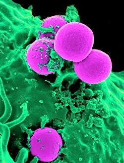
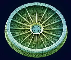
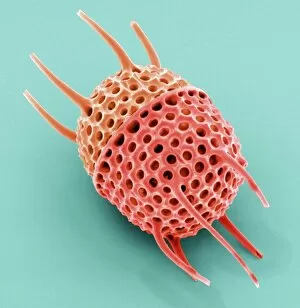
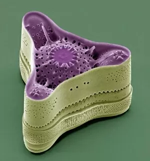
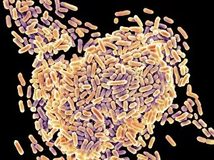
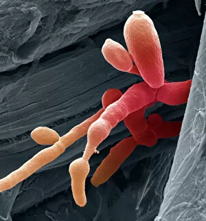
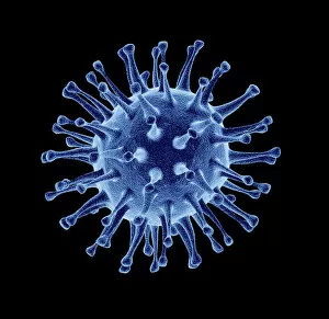
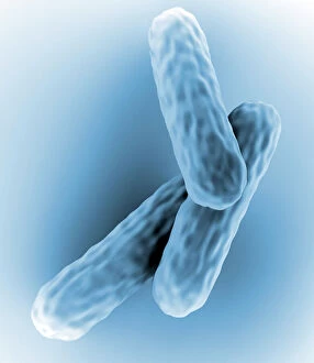
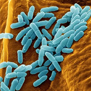
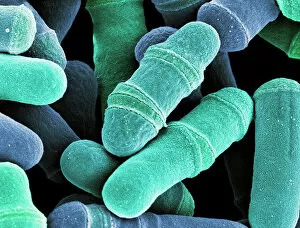
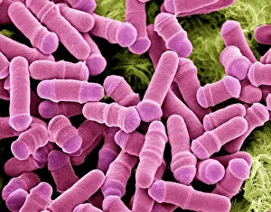
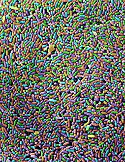
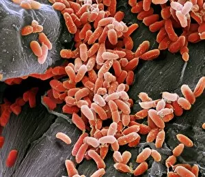
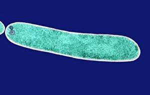
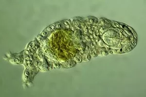
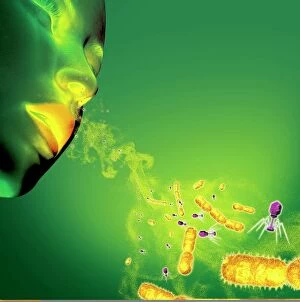
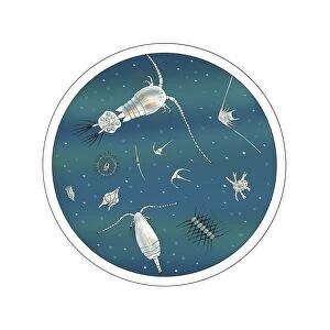
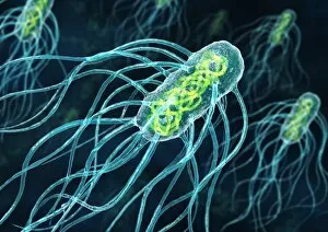
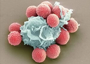
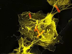
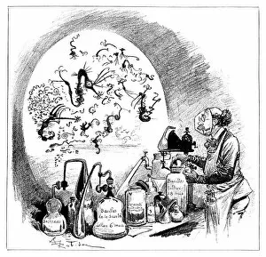
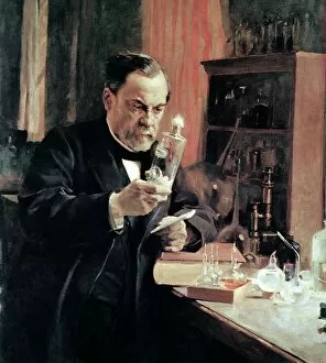
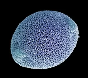
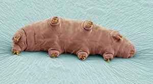
Microbiological wonders unfold in this captivating collection of images. From the delicate budding yeast cell to the intricate calcareous phytoplankton, every specimen reveals the beauty and complexity of the microscopic world. The avian flu virus, with its menacing structure, serves as a reminder of nature's power. Meanwhile, a neutrophil engulfing MRSA showcases our immune system's remarkable defense mechanisms. E. Coli and Salmonella bacteria take center stage under the scanning electron microscope, their distinct shapes and patterns mesmerizing to behold. Diatoms enchant with their ornate structures, resembling tiny jewels adorning aquatic landscapes. Intriguingly diverse forms continue to amaze as we encounter Candida fungus in all its textured glory. The HIV particle appears both mysterious and formidable—a silent invader that has challenged humanity for decades. Artistic renditions depict various cell types, offering a visual representation of life at its most fundamental level. Finally, historical diagrams showcasing anthrax cultures remind us of our ongoing battle against infectious diseases throughout history. These microbiological glimpses invite us into an unseen realm teeming with lifeforms that shape our world in ways we may never fully comprehend.